 Back
BackThe Nervous System: Structure, Function, and Physiology
Study Guide - Smart Notes
 Tailored notes based on your materials, expanded with key definitions, examples, and context.
The Nervous System: Overview
Main Divisions of the Nervous System
The nervous system is responsible for coordinating and integrating all bodily functions through rapid communication. It is divided into three main divisions: the Central Nervous System (CNS), the Peripheral Nervous System (PNS), and the Enteric Nervous System (ENS).
Central Nervous System (CNS): Consists of the brain and spinal cord. It is the main center for information processing, integration, and coordination.
Peripheral Nervous System (PNS): Includes all neural tissue outside the CNS. It is subdivided into sensory (afferent) and motor (efferent) divisions.
Enteric Nervous System (ENS): Embedded in the wall of the gastrointestinal tract, it helps control digestive function (covered in more detail in A&P II).
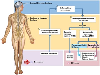
Functional Organization of the PNS
Sensory (Afferent) Division: Transmits information from sensory receptors to the CNS.
Motor (Efferent) Division: Carries commands from the CNS to effector organs (muscles and glands). It is further divided into:
Somatic Nervous System (SNS): Controls voluntary movements of skeletal muscles.
Autonomic Nervous System (ANS): Regulates involuntary functions (smooth muscle, cardiac muscle, glands, adipose tissue). The ANS is subdivided into the sympathetic and parasympathetic divisions.
Cells of the Nervous System
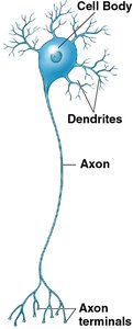
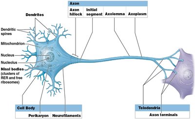
Neurons: Structure and Function
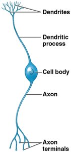
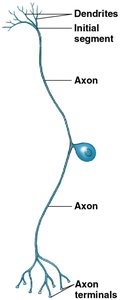
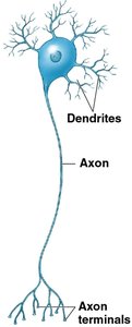
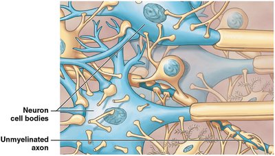
Neurons are the primary cells responsible for communication within the nervous system. They are typically large, highly specialized, and rarely divide after development. Neurons have a unique structure that supports their function in transmitting electrical and chemical signals.
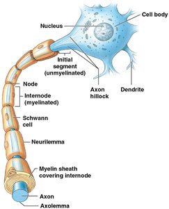
Cell Body (Soma): Contains the nucleus and organelles; responsible for metabolic activities.
Dendrites: Branch-like extensions that receive signals from other neurons.
Axon: A long projection that conducts electrical impulses away from the cell body toward other neurons or effectors.
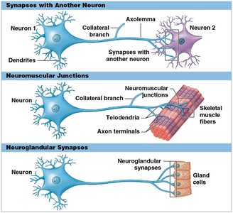
Axon Terminals: The endpoints where neurotransmitters are released to communicate with other cells.
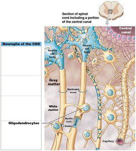
Neuroglial Cells (Glia)
Neuroglia are support cells that provide structural and metabolic support to neurons. They are smaller than neurons and can divide throughout life. Types include:
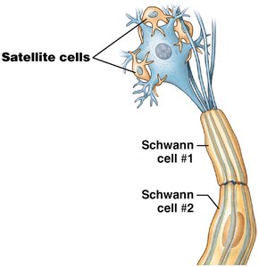
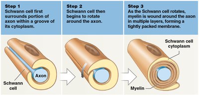
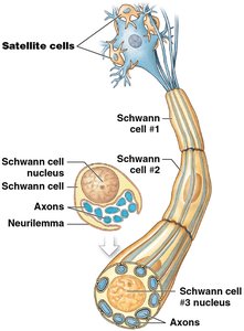
Schwann Cells: Form myelin in the PNS and assist in axon repair.
Satellite Cells: Surround neuron cell bodies in the PNS, regulating the environment.
Microglia: Phagocytic cells that remove waste and pathogens in the CNS.
Astrocytes: Maintain the blood-brain barrier, regulate interstitial fluid, and form scar tissue after injury.
Ependymal Cells: Line ventricles and produce cerebrospinal fluid (CSF).
Oligodendrocytes: Form myelin in the CNS.
Neuron Classification
Structural and Functional Types
Neurons can be classified based on their structure and function:
Structural Types:
Anaxonic Neurons: No obvious axon; found in the brain and special sense organs.
Bipolar Neurons: One dendrite and one axon; found in sensory organs (e.g., retina).
Unipolar Neurons: Single process with cell body off to the side; most sensory neurons in the PNS.
Multipolar Neurons: Many dendrites, one axon; most common type in the CNS.
Functional Types: Sensory (afferent), Motor (efferent), and Interneurons (association neurons).
Neuroglia and Myelination
Neuroglia of the CNS
Neuroglia in the CNS include astrocytes, microglia, ependymal cells, and oligodendrocytes. Oligodendrocytes are responsible for forming myelin sheaths around axons, which increases the speed of neural transmission.
White Matter: Composed of myelinated axons; appears white due to lipid content.
Gray Matter: Contains neuron cell bodies and unmyelinated axons.
Neuroglia of the PNS
Schwann Cells: Form myelin sheaths in the PNS and assist in axon repair.
Satellite Cells: Surround neuron cell bodies in the PNS and regulate the environment.
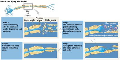
Axon Repair in the PNS
Schwann cells play a crucial role in the repair of damaged axons in the PNS. After injury, Schwann cells proliferate and form a regeneration tube that guides the regrowth of the axon.
Neuronal Physiology
Resting Membrane Potential
Neurons maintain a resting membrane potential of approximately -70 mV, primarily due to the activity of the sodium-potassium pump, which moves 3 Na+ ions out and 2 K+ ions into the cell, creating a charge differential across the membrane.
Key Players: Sodium-potassium pump, selective permeability, and large negatively charged proteins inside the cell.
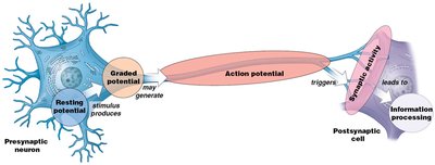
Graded Potentials
Graded potentials are changes in membrane potential that vary in size and are produced by ligand-gated channels in response to neurotransmitter binding. If the graded potential is large enough to reach threshold (-55 mV), it can trigger an action potential.
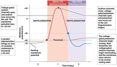
Action Potentials
Action potentials are rapid, all-or-none electrical impulses generated by voltage-gated channels. They propagate along the axon and are essential for neural communication.
Depolarization: Na+ channels open, Na+ enters the cell, making the inside more positive.
Repolarization: K+ channels open, K+ leaves the cell, restoring negativity.
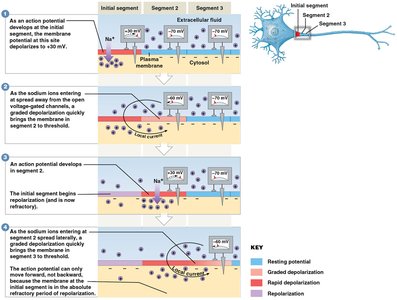
Refractory Period: Time during which a neuron cannot fire another action potential.
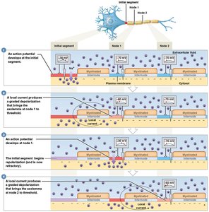
Propagation of Action Potentials
Action potentials can propagate in two ways:
Continuous Propagation: Occurs in unmyelinated axons; the action potential moves along every segment of the membrane.
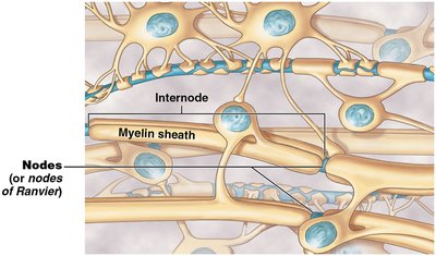
Saltatory Conduction: Occurs in myelinated axons; the action potential jumps from node to node (Nodes of Ranvier), increasing speed.
Synaptic Activity
Structure and Function of Synapses
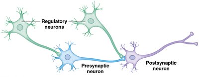
A synapse is the site of communication between two neurons or between a neuron and an effector cell. The presynaptic neuron releases neurotransmitters that cross the synaptic cleft and bind to receptors on the postsynaptic cell, generating a response.
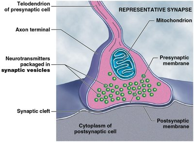
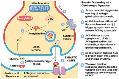
Events at a Cholinergic Synapse
Action potential arrives at the axon terminal, opening voltage-gated calcium channels.
Calcium influx triggers exocytosis of acetylcholine (ACh) into the synaptic cleft.
ACh binds to receptors on the postsynaptic membrane, opening sodium channels and generating a graded potential.
ACh is broken down by acetylcholinesterase (AChE), ending the signal.
Choline is reabsorbed and recycled by the presynaptic neuron.
Excitatory and Inhibitory Synapses
Not all neurotransmitters are excitatory. Some open potassium channels, causing hyperpolarization (inhibitory postsynaptic potentials, IPSPs), while others open sodium channels, causing depolarization (excitatory postsynaptic potentials, EPSPs). The net effect of all inputs determines whether the postsynaptic neuron will fire an action potential.
Summary Table: Types of Neurons and Neuroglia
Cell Type | Location | Main Function |
|---|---|---|
Neuron | CNS & PNS | Transmit electrical and chemical signals |
Astrocyte | CNS | Blood-brain barrier, regulate environment |
Oligodendrocyte | CNS | Form myelin sheaths |
Microglia | CNS | Phagocytosis of debris/pathogens |
Ependymal cell | CNS | Produce cerebrospinal fluid |
Schwann cell | PNS | Form myelin, assist in repair |
Satellite cell | PNS | Regulate environment around neurons |
Additional info: This guide covers the structure and function of the nervous system, neuron physiology, neuroglia, and synaptic activity, providing a foundation for further study in anatomy and physiology.
