 Back
BackCh 12: The Nervous System: Structure, Function, and Neural Activity
Study Guide - Smart Notes
 Tailored notes based on your materials, expanded with key definitions, examples, and context.
An Introduction to the Nervous System
Overview of the Nervous System
The nervous system is responsible for coordinating all activities in the body by transmitting signals between different parts. It consists of specialized cells called neurons and supporting cells known as neuroglia (or glial cells). The main organs include the brain, spinal cord, peripheral nerves, cranial nerves, and special sense organs.
Neurons: Cells that send and receive electrical and chemical signals.
Neuroglia: Cells that support, protect, and nourish neurons.
Divisions of the Nervous System
Anatomical Divisions
The nervous system is divided into two main anatomical parts:
Central Nervous System (CNS): Composed of the brain and spinal cord. It processes information and coordinates activity.
Peripheral Nervous System (PNS): Includes all neural tissue outside the CNS, such as peripheral nerves and ganglia.
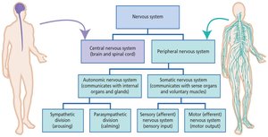
Functional Divisions of the PNS
The PNS is further divided based on function:
Somatic Nervous System (SNS): Controls voluntary movements and transmits sensory information.
Autonomic Nervous System (ANS): Regulates involuntary functions (e.g., heart rate, digestion). Subdivided into:
Sympathetic Division: Arousing responses (fight or flight).
Parasympathetic Division: Calming responses (rest and digest).
Neurons: Structure and Function
Basic Structure of Neurons
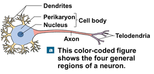
Neurons are the basic functional units of the nervous system. The most common type in the CNS is the multipolar neuron, which has a cell body, multiple dendrites, and a single long axon.
Cell Body (Soma): Contains the nucleus and organelles.
Dendrites: Highly branched extensions that receive signals from other neurons.
Axon: Long process that transmits electrical impulses away from the cell body.
Telodendria: Fine extensions at the end of the axon.
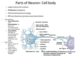
Key Terms for Axon Structure
Axoplasm: Cytoplasm of the axon, containing neurofibrils, neurotubules, enzymes, and organelles.
Axolemma: Specialized cell membrane covering the axoplasm.
Axon Hillock: Thickened region of the cell body where the axon originates; site of action potential initiation.
Collaterals: Branches of a single axon.
Synaptic Terminals: Tips of telodendria where neurotransmitters are released.
The Synapse and Neurotransmission
Structure of the Synapse
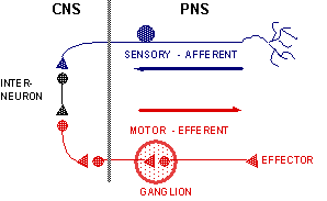
A synapse is the junction where a neuron communicates with another cell (neuron, muscle, or gland). It consists of:
Presynaptic Cell: Neuron sending the signal.
Postsynaptic Cell: Cell receiving the signal.
Synaptic Cleft: Small gap between the presynaptic and postsynaptic membranes.
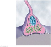
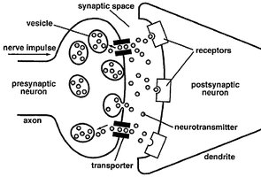
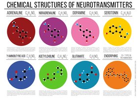
Neurotransmitters
Neurotransmitters are chemical messengers released from the presynaptic terminal. They bind to receptors on the postsynaptic membrane, causing changes in the postsynaptic cell. They are broken down by enzymes and can be reassembled at the synaptic terminal.
Norepinephrine (NE): Sympathetic neurotransmitter.
Acetylcholine (ACh): Parasympathetic and somatic motor neurotransmitter.
Dopamine: Basal ganglia neurotransmitter.
GABA: Inhibitory neurotransmitter in the cortex.
Glutamate: Involved in learning and memory.
Serotonin: Mood regulation; used by neurons in the midbrain, pons, and medulla.
Classification of Neurons
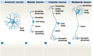
Structural Types of Neurons
Neurons are classified based on their structure:
Anaxonic Neurons: Found in the brain and sense organs; no obvious axon.
Bipolar Neurons: Found in special sensory organs (sight, smell, hearing); one dendrite and one axon.
Unipolar Neurons: Found in sensory neurons of the PNS; single process splits into two branches.
Multipolar Neurons: Common in the CNS; multiple dendrites and one axon.
Neuroglia (Glial Cells)
Types and Functions of Neuroglia
Neuroglia are supporting cells in the nervous system. They are essential for neuron function, repair, and maintenance.
CNS Neuroglia:
Ependymal Cells: Line ventricles; produce and circulate cerebrospinal fluid.
Astrocytes: Maintain blood-brain barrier; provide structural support.
Oligodendrocytes: Form myelin sheaths in the CNS.
Microglia: Act as phagocytes; remove debris and pathogens.
PNS Neuroglia:
Satellite Cells: Surround neuron cell bodies in ganglia; regulate environment.
Schwann Cells: Form myelin sheaths around peripheral axons; aid in regeneration.
Neural Responses to Injury
PNS Neuron Repair and Regeneration
Peripheral neurons can regenerate after injury, primarily due to the presence of Schwann cells. The process involves:
Wallerian Degeneration: Distal axon degenerates after injury.
Schwann Cells: Form a path for new axon growth and wrap the new axon in myelin.
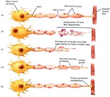
Additional info: CNS neurons have limited regeneration due to the presence of oligodendrocytes and inhibitory factors.
Membrane Potentials and Neural Activity
Five Main Membrane Processes
Neural activity depends on changes in membrane potential, which include:
Resting Potential: The baseline transmembrane potential of a resting cell.
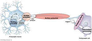
Graded Potential: Temporary, localized change in resting potential caused by a stimulus.
Action Potential: Electrical impulse that propagates along the axon.
Synaptic Activity: Release of neurotransmitters at the synapse, producing graded potentials in the postsynaptic cell.
Information Processing: Integration of stimuli by the postsynaptic cell.
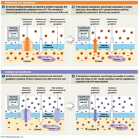
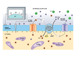
Resting Membrane Potential
The resting membrane potential is established by passive forces across the membrane, including chemical and electrical gradients of ions (mainly Na+ and K+).
Chemical Gradients: Differences in ion concentration across the membrane.
Electrical Gradients: Separation of charges across the membrane, creating a potential difference.
Membrane Potential Terminology
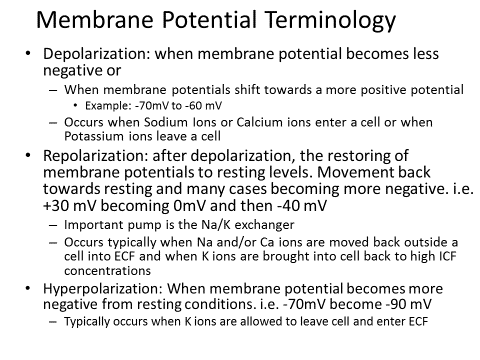
Depolarization: Membrane potential becomes less negative (e.g., -70 mV to -60 mV); occurs when Na+ or Ca2+ enters the cell or K+ leaves.
Repolarization: Return to resting potential after depolarization; typically involves K+ leaving the cell.
Hyperpolarization: Membrane potential becomes more negative than resting (e.g., -70 mV to -90 mV); usually due to K+ leaving the cell.
Graded Potentials
Characteristics of Graded Potentials
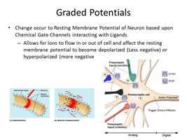
Graded potentials are changes in membrane potential that vary in size and decrease with distance from the stimulus. They can be depolarizing or hyperpolarizing.
Most change occurs at the site of stimulation; effect decreases with distance.
Spread passively due to local currents.
May involve depolarization or hyperpolarization.
Stronger stimulus produces greater change and affects a larger area.
Action Potentials
Generation and Propagation
An action potential is a rapid, all-or-none electrical event triggered when the membrane potential reaches a threshold. The process involves:
Depolarization to Threshold: Stimulus causes membrane potential to reach threshold.
Activation of Na+ Channels: Rapid influx of sodium ions causes further depolarization.
Inactivation of Na+ Channels and Activation of K+ Channels: Sodium channels close, potassium channels open, leading to repolarization.
Return to Normal Permeability: Potassium channels close, membrane returns to resting potential.
Refractory Periods:
Absolute Refractory Period: No action potential can be generated.
Relative Refractory Period: Action potential possible only with a stronger stimulus.
Factors Affecting Conduction Velocity
Axon Diameter: Larger diameter = lower resistance = faster conduction.
Myelination: Myelinated axons conduct impulses faster.
Types of Axons
Type | Myelination | Diameter | Speed | Function |
|---|---|---|---|---|
Type A | Myelinated | Large | 140 m/sec | Rapid information (position, balance, touch, motor) |
Type B | Myelinated | Medium | 18 m/sec | Intermediate signals (sensory, peripheral effectors) |
Type C | Unmyelinated | Small | 1 m/sec | Slow information (involuntary muscle, glands) |
Neuromodulators
Definition and Examples
Neuromodulators are molecules that modulate the activity of neurons, often by acting on G-protein coupled receptors. They can increase or decrease the effect of neurotransmitters and may remain in the cerebrospinal fluid for extended periods.
Opioids: Block pain pathways, reduce norepinephrine turnover, increase dopamine renewal.
Cocaine: Blocks dopamine reuptake, increasing dopamine activity and inducing pleasure.
Information Processing in Neurons
Postsynaptic Potentials and Summation
Postsynaptic potentials are graded changes in the postsynaptic cell membrane potential in response to neurotransmitters.
Excitatory Postsynaptic Potential (EPSP): Graded depolarization, increases likelihood of action potential.
Inhibitory Postsynaptic Potential (IPSP): Graded hyperpolarization, decreases likelihood of action potential.
Summation: EPSPs and IPSPs combine at the axon hillock. Types include:
Temporal Summation: Rapid, repeated stimulation by one neuron.
Spatial Summation: Simultaneous stimulation by multiple neurons.
Summary and Review
Neurons are the functional cells of the nervous system, supported by neuroglia.
Resting membrane potential, graded potentials, and action potentials are key to neural signaling.
Neurotransmitters and synapses enable communication between neurons.
Conduction velocity is influenced by axon diameter and myelination.
