 Back
BackThe Nervous System: Structure, Function, and Organization
Study Guide - Smart Notes
 Tailored notes based on your materials, expanded with key definitions, examples, and context.
Nervous System Overview
Introduction to the Nervous System
The nervous system is the primary control and communication system of the body. It monitors both external and internal environments, regulates vital body functions such as heart rate and temperature, and coordinates movement, behavior, thoughts, and emotions.
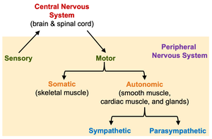
Central Nervous System (CNS): The command center, consisting of the brain and spinal cord. It processes sensory input and directs motor responses.
Peripheral Nervous System (PNS): All nervous tissue outside the CNS, including cranial and spinal nerves. It transmits signals between the CNS and the rest of the body.
Functional Organization
Sensory (Afferent) Division: Transmits sensory information from receptors to the CNS.
Motor (Efferent) Division: Carries commands from the CNS to effector organs (muscles and glands).
Somatic Nervous System: Controls voluntary movements via skeletal muscles.
Autonomic Nervous System: Regulates involuntary functions (smooth muscle, cardiac muscle, glands). Subdivided into:
Sympathetic Division: Mobilizes body systems during activity (fight or flight).
Parasympathetic Division: Conserves energy and promotes maintenance functions (rest and digest).
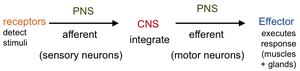
Pathway of Nervous System Function
The nervous system operates through a sequence of detection, integration, and response:
Receptors detect stimuli.
Sensory (afferent) neurons relay information to the CNS.
Integration occurs in the CNS.
Motor (efferent) neurons carry commands to effectors (muscles/glands).
Histology of the Nervous System
Neurons
Neurons are the functional cells of the nervous system, specialized for generating and conducting electrical signals. Most are amitotic (do not divide), with exceptions in taste, olfaction, and memory.
Cell Body: Contains the nucleus and organelles. Clusters in CNS are called nuclei; in PNS, ganglia.
Dendrites: Receive signals and transmit them to the cell body.
Axon: Conducts signals away from the cell body. May be myelinated (increases conduction speed) or unmyelinated.
Myelin Sheath: Formed by Schwann cells (PNS) or oligodendrocytes (CNS); interrupted by nodes of Ranvier.
Neuroglia (Glial Cells)
Supportive cells that protect, nourish, and insulate neurons. They are mitotic and can give rise to tumors.
CNS Glia: Oligodendrocytes (myelination), astrocytes (blood-brain barrier), microglia (immune defense), ependymal cells (produce/circulate CSF).
PNS Glia: Schwann cells (myelination), satellite cells (support neuron cell bodies in ganglia).
Neuron Classification
Structural Types:
Unipolar: One process; always sensory.
Bipolar: Two processes; sensory (e.g., retina, olfaction).
Multipolar: One axon, multiple dendrites; most common, includes all motor and interneurons.
Functional Types:
Sensory (Afferent): Transmit signals to CNS; mostly unipolar.
Interneurons: Integrate information within CNS; multipolar.
Motor (Efferent): Transmit signals from CNS to effectors; multipolar.
Synapses
Neuronal Junction: Neuron to neuron.
Neuromuscular Junction: Neuron to skeletal muscle.
Neuroglandular Junction: Neuron to gland.
Chemical Synapse: Neurotransmitter released from presynaptic neuron crosses synaptic cleft to postsynaptic neuron, generating a new electrical signal.
Central Nervous System: Brain
Cerebrum
The largest part of the brain, divided into right and left hemispheres and further into lobes (frontal, temporal, parietal, occipital, insula). Responsible for higher brain functions, including movement, sensation, memory, and intellect.
Surface Features: Fissures (deep grooves), gyri (ridges), sulci (shallow grooves).
Cerebral Cortex: Gray matter; site of conscious mind, divided into motor, sensory, and association areas.
White Matter: Myelinated tracts connecting different brain regions.
Basal Nuclei: Masses of gray matter involved in movement regulation.
Other Major Brain Regions
Thalamus: Relay station for sensory information to the cortex.
Hypothalamus: Regulates autonomic functions (e.g., heart rate, temperature).
Brainstem: Includes midbrain (reflexes), pons (respiratory centers), and medulla (vital centers for heart, vessels, breathing).
Cerebellum: Coordinates movement, balance, and posture.
Functional Systems
Limbic System: Regulates emotions and memory.
Reticular Activating System (RAS): Controls alertness and consciousness.
Central Nervous System: Spinal Cord
Functions and Structure
Acts as a communication highway between brain and body.
Coordinates reflexes for rapid responses.
Extends from foramen magnum to L1/L2, ending at the conus medullaris; cauda equina are nerve roots extending below.
Gray Matter: H-shaped; contains cell bodies and dendrites.
White Matter: Myelinated axons forming ascending (sensory) and descending (motor) tracts.
Central Nervous System: Coverings and Support
Meninges
Dura Mater: Outermost layer; two layers in brain, one in spinal cord.
Arachnoid Mater: Middle layer; contains CSF in subarachnoid space.
Pia Mater: Innermost, vascular layer attached to CNS surface.
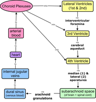
Cerebrospinal Fluid (CSF)
CSF is an extracellular fluid that cushions the brain and spinal cord, produced by choroid plexuses in the ventricles. It circulates through the ventricles, central canal, and subarachnoid space, and is reabsorbed into the blood via arachnoid granulations.
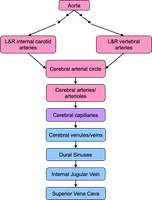
Central Nervous System: Blood Supply
Cerebral Arterial Circle (Circle of Willis)
A circular arrangement of arteries at the base of the brain that ensures continuous blood supply even if one part is blocked. Formed by the joining of internal carotid and vertebral arteries.
Blood Brain Barrier (BBB)
Formed by tight junctions between capillary endothelial cells and astrocyte foot processes.
Regulates passage of substances from blood to CNS, protecting the brain from toxins and pathogens.
Allows passage of glucose, O2, CO2, alcohol, and some drugs; blocks cells and many drugs.
Peripheral Nervous System: Structure and Function
Cranial and Spinal Nerves
Cranial Nerves: 12 pairs; sensory, motor, or mixed.
Spinal Nerves: 31 pairs; all mixed nerves, exit via intervertebral foramina, and form plexuses (except thoracic nerves T2-T12).
Nerve Structure
Epineurium: Surrounds entire nerve.
Perineurium: Surrounds fascicles (bundles of axons).
Endoneurium: Surrounds individual axons.
Peripheral Nervous System: Sensory (Afferent) Division
Sensory Receptors
By Location: Exteroceptors (external), interoceptors (internal), proprioceptors (body position).
By Stimulus Type: Mechanoreceptors (touch, pressure), thermoreceptors (temperature), chemoreceptors (chemicals), photoreceptors (light), nociceptors (pain).
By Structure: Free nerve endings, encapsulated nerve endings.
Peripheral Nervous System: Motor (Efferent) Division
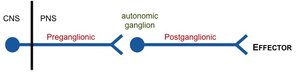
Somatic vs. Autonomic Nervous System
Somatic: Single multipolar neuron from CNS to skeletal muscle.
Autonomic: Two-neuron chain (preganglionic and postganglionic) from CNS to smooth/cardiac muscle or glands.
Sympathetic Division: "Fight or flight"; preganglionic neurons in thoracic/lumbar spinal cord, ganglia near spinal cord, long postganglionic axons.
Parasympathetic Division: "Rest and digest"; preganglionic neurons in brainstem/sacral spinal cord, ganglia near/in effector, short postganglionic axons.
Nervous System Pathways
Ascending (Sensory) Pathways
Three-neuron chain: first-order (receptor to CNS), second-order (spinal cord/medulla to thalamus), third-order (thalamus to cortex).
Example: Dorsal column pathway for touch.
Descending (Motor) Pathways
Upper motor neuron (cortex/brainstem to spinal cord), lower motor neuron (spinal cord to muscle).
Direct (corticospinal) tracts control voluntary movement; indirect tracts regulate posture and muscle tone.
Related Medical Conditions
Meningitis: Inflammation of the meninges, usually due to infection.
Neuropathy: Damage to peripheral nerves (e.g., carpal tunnel syndrome, Bell’s palsy).
