 Back
The Nervous System: Structure, Function, and Organization
Study Guide - Smart Notes
 Tailored notes based on your materials, expanded with key definitions, examples, and context.
The Nervous System: Overview
Introduction to the Nervous System
The nervous system is the master controlling and communicating system of the body. It utilizes electrical and chemical signals to coordinate rapid and specific responses, ensuring immediate reactions to internal and external stimuli.
Sensory Input: Information gathered by sensory receptors about changes inside and outside the body.
Integration: Processing and interpretation of sensory input to determine an appropriate response.
Motor Output: Activation of effector organs (muscles and glands) to produce a response.
Divisions of the Nervous System
Central and Peripheral Nervous Systems
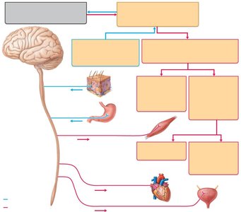
The nervous system is divided into the central nervous system (CNS) and the peripheral nervous system (PNS), each with distinct structures and functions.
Central Nervous System (CNS): Consists of the brain and spinal cord; serves as the integration and control center.
Peripheral Nervous System (PNS): Composed of nerves extending from the brain and spinal cord, including cranial and spinal nerves. It acts as communication lines between the CNS and the rest of the body.
Functional Divisions of the PNS
Sensory (Afferent) Division: Transmits impulses from sensory receptors to the CNS. Includes somatic sensory fibers (from skin, muscles, joints) and visceral sensory fibers (from organs).
Motor (Efferent) Division: Transmits impulses from the CNS to effector organs. Subdivided into:
Somatic Nervous System: Voluntary control of skeletal muscles.
Autonomic Nervous System (ANS): Involuntary control of smooth muscle, cardiac muscle, and glands. Includes sympathetic and parasympathetic divisions, which generally have opposing effects.
Histology of Nervous Tissue
Neuroglia (Glial Cells)
Nervous tissue is highly cellular, with two principal cell types: neuroglia and neurons. Neuroglia support and protect neurons, while neurons transmit electrical signals.
Astrocytes: Most abundant glial cells in the CNS; support neurons, regulate the chemical environment, and participate in information processing.
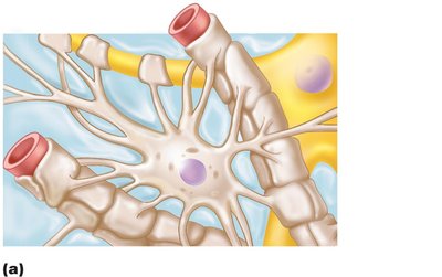
Microglial Cells: Small, defensive cells that monitor neuron health and can phagocytize debris and pathogens.
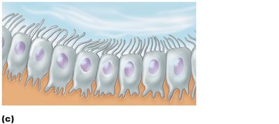
Ependymal Cells: Line the central cavities of the brain and spinal cord; may be ciliated to help circulate cerebrospinal fluid (CSF).
Oligodendrocytes: Form myelin sheaths around CNS nerve fibers, providing electrical insulation.
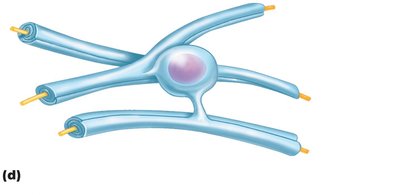
Satellite Cells (PNS): Surround neuron cell bodies in the PNS, similar in function to astrocytes.
Schwann Cells (PNS): Form myelin sheaths around peripheral nerve fibers and assist in nerve regeneration.
Neurons (Nerve Cells)
Neurons are the structural and functional units of the nervous system. They are specialized for impulse conduction, have extreme longevity, are mostly amitotic, and have a high metabolic rate.
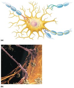
Cell Body (Soma): Contains the nucleus and organelles; biosynthetic center of the neuron.
Dendrites: Short, branched processes that receive input and convey signals toward the cell body.
Axon: Single, long process that transmits impulses away from the cell body to other neurons or effectors.
Myelin Sheath
Structure and Function
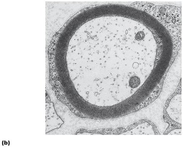
The myelin sheath is a segmented, whitish, protein-lipoid covering that insulates axons, increasing the speed of nerve impulse transmission. Myelinated fibers conduct impulses rapidly, while nonmyelinated fibers conduct more slowly.
PNS Myelination: Schwann cells wrap around axons, forming the myelin sheath. Gaps between Schwann cells are called nodes of Ranvier.
CNS Myelination: Oligodendrocytes form myelin sheaths around multiple axons. White matter consists of myelinated fibers; gray matter contains neuron cell bodies and unmyelinated fibers.
Classification of Neurons
Structural Classification
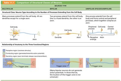
Neurons are classified by the number of processes extending from the cell body:
Type | Structure | Location |
|---|---|---|
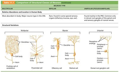
Multipolar | Many dendrites, one axon | Most abundant; CNS |
Bipolar | One dendrite, one axon | Special senses (retina, olfactory mucosa) |
Unipolar | Single process splits into peripheral and central branches | PNS (dorsal root ganglia, sensory ganglia of cranial nerves) |
Functional Classification
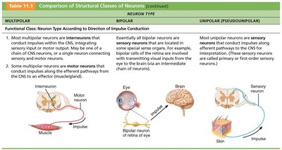
Sensory (Afferent) Neurons: Transmit impulses from sensory receptors toward the CNS; mostly unipolar.
Motor (Efferent) Neurons: Carry impulses from the CNS to effectors; multipolar.
Interneurons: Lie between sensory and motor neurons; shuttle signals through CNS pathways; most abundant.
Membrane Potentials and Neural Signaling
Membrane Potential Changes
Neurons use changes in membrane potential as communication signals. These changes occur when ion concentrations or membrane permeability change.
Graded Potentials: Short-distance signals, usually in dendrites or cell bodies.
Action Potentials: Long-distance signals generated in axons; involve a rapid reversal of membrane potential (~100 mV).
Depolarization: Membrane potential becomes less negative, increasing the likelihood of a nerve impulse.
Hyperpolarization: Membrane potential becomes more negative, reducing the likelihood of a nerve impulse.
The Synapse
Structure and Function
Synapses are junctions that mediate information transfer between neurons or between a neuron and an effector cell. They can be classified by their anatomical location:
Axodendritic: Between axon terminals and dendrites.
Axosomatic: Between axon terminals and neuron cell bodies.
Other types: Axoaxonic, dendrodendritic, somatodendritic.
Presynaptic Neuron: Conducts impulses toward the synapse.
Postsynaptic Neuron: Transmits impulses away from the synapse.
Central Nervous System: Brain Structure and Function
Major Brain Regions
Cerebral Hemispheres: Frontal, parietal, temporal, occipital, and insula lobes.
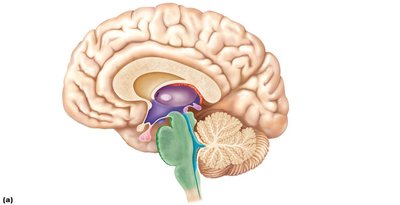
Diencephalon: Thalamus, hypothalamus, epithalamus.
Brain Stem: Midbrain, pons, medulla oblongata.
Cerebellum: Coordinates movement and balance.
Cerebral Cortex
The cerebral cortex is the site of conscious mind functions, including awareness, sensory perception, voluntary motor initiation, communication, memory, and understanding. It is organized into motor, sensory, and association areas.
Primary Motor Cortex: Controls voluntary movements; represented by the motor homunculus.
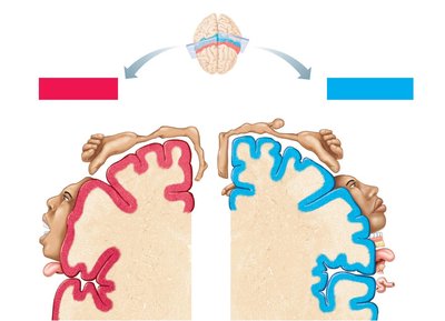
Premotor Cortex: Plans and coordinates complex movements.
Prefrontal Cortex: Involved in intellect, cognition, personality, and decision-making.
Posterior Association Area: Involved in pattern recognition, spatial localization, and language comprehension.
Limbic Association Area: Provides emotional context and memory formation.
Diencephalon
Thalamus: Gateway to the cerebral cortex; sorts, edits, and relays sensory information.
Hypothalamus: Regulates autonomic functions, emotions, body temperature, hunger, thirst, sleep-wake cycles, and endocrine activity.
Epithalamus: Contains the pineal gland, which secretes melatonin to regulate sleep-wake cycles.
Peripheral Nervous System: Structure and Function
Sensory Receptors and Nerves
The PNS links the CNS to the external environment through sensory receptors and nerves. Sensory receptors are classified by stimulus type, location, and structural complexity.
Mechanoreceptors: Respond to touch, pressure, vibration, and stretch.
Thermoreceptors: Detect temperature changes.
Photoreceptors: Respond to light (e.g., in the retina).
Chemoreceptors: Detect chemicals (e.g., taste, smell, blood chemistry).
Nociceptors: Respond to pain-causing stimuli.
Classification of Nerves
Mixed Nerves: Contain both sensory and motor fibers.
Sensory (Afferent) Nerves: Carry impulses toward the CNS.
Motor (Efferent) Nerves: Carry impulses away from the CNS.
Regeneration and Reflexes
Regeneration of Nerve Fibers
Peripheral axons can regenerate if the cell body is intact, but CNS fibers rarely regenerate due to inhibitory factors and scar tissue formation.
PNS Regeneration: Involves Wallerian degeneration, macrophage cleanup, and axon regrowth through a regeneration tube formed by Schwann cells.
CNS Regeneration: Inhibited by oligodendrocyte proteins and astrocyte scar tissue.
Reflexes and Reflex Arcs
Reflexes are rapid, involuntary responses to stimuli. Reflex arcs consist of five components: receptor, sensory neuron, integration center, motor neuron, and effector.
Inborn Reflexes: Present at birth; maintain posture and control visceral activities.
Learned Reflexes: Acquired through practice (e.g., driving skills).
Autonomic Nervous System (ANS)
Structure and Function
The ANS regulates involuntary functions by innervating smooth muscle, cardiac muscle, and glands. It operates subconsciously and is divided into sympathetic and parasympathetic divisions, which generally have antagonistic effects to maintain homeostasis.
Sympathetic Division: Mobilizes the body during activity ("fight-or-flight").
Parasympathetic Division: Promotes maintenance activities and conserves energy ("rest-and-digest").
Effects of Drugs on the ANS
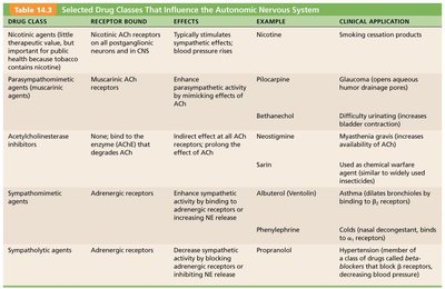
Certain drugs can influence the ANS by acting on specific receptors or enzymes. These drugs are used clinically to treat various conditions.
Drug Class | Receptor Bound | Effects | Example | Clinical Application |
|---|---|---|---|---|
Nicotinic agents | Nicotinic ACh receptors | Stimulate all postganglionic neurons; increase blood pressure | Nicotine | Smoking cessation products |
Parasympathomimetic agents | Muscarinic ACh receptors | Enhance parasympathetic activity | Pilocarpine | Glaucoma (opens aqueous humor drainage pores) |
Acetylcholinesterase inhibitors | Enzyme AChE | Indirect effect at all ACh receptors; prolong ACh effect | Neostigmine | Myasthenia gravis (increases availability of ACh) |
Sympathomimetic agents | Adrenergic receptors | Enhance sympathetic activity | Albuterol | Asthma (dilates bronchioles) |
Sympathetic agents | Adrenergic receptors | Decrease sympathetic activity | Propranolol | Hypertension (decreases blood pressure) |
Developmental Aspects of the ANS
Aging affects the ANS, leading to conditions such as constipation, dry eyes, frequent eye infections, and orthostatic hypotension due to less responsive pressure receptors and declining cardiovascular control.
