 Back
BackThe Nervous System: Structure, Function, and Physiology
Study Guide - Smart Notes
 Tailored notes based on your materials, expanded with key definitions, examples, and context.
The Nervous System
Introduction to the Nervous System
The nervous system is one of the primary organ systems responsible for maintaining homeostasis by coordinating the activities of the body. It works closely with the endocrine system, but is distinguished by its rapid and brief responses. The nervous system is the most complex organ system in the body.
Key Functions:
Monitors internal and external environments
Integrates sensory information
Coordinates voluntary and involuntary responses

Anatomical Divisions of the Nervous System
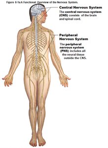
Central Nervous System (CNS): Consists of the brain and spinal cord. Responsible for integrating and coordinating sensory processing and motor output. Site of higher functions such as intelligence, memory, and emotion.
Peripheral Nervous System (PNS): Includes all neural tissue outside the CNS. Acts as a communication link between the CNS and the rest of the body.
Functional Divisions of the PNS
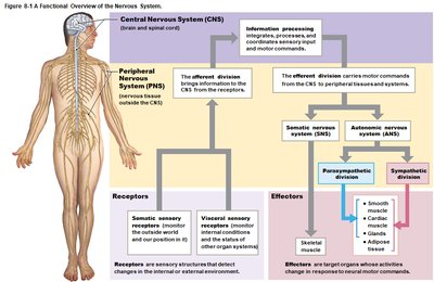
Afferent Division: Brings sensory information to the CNS from receptors in tissues and organs.
Efferent Division: Carries motor commands from the CNS to effectors (muscles and glands).
Effectors: Target organs whose activities change in response to neural commands.
Subdivisions of the Efferent Division
Somatic Nervous System (SNS): Controls skeletal muscle contractions (voluntary and involuntary).
Autonomic Nervous System (ANS): Controls smooth muscle, cardiac muscle, and glands. Subdivided into:
Sympathetic Division (fight-or-flight)
Parasympathetic Division (rest-and-digest)
Enteric Nervous System (ENS): Controls digestive tract functions
Nervous Tissue
Types of Cells in Nervous Tissue
Neurons: Basic functional units of the nervous system. Specialized for communication.
Neuroglia (Glial Cells): Support, protect, and nourish neurons. Capable of cell division.
Structure of a Typical Neuron
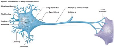
Neurons have a unique structure that supports their function in communication.
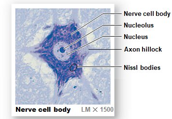
Cell Body (Soma): Contains the nucleus, nucleolus, mitochondria, Nissl bodies (RER and free ribosomes), and axon hillock (origin of action potentials).
Dendrites: Receive incoming signals from other neurons.
Axon: Transmits electrical impulses away from the cell body toward other cells.
Axon Terminals: Release neurotransmitters to communicate with other cells.
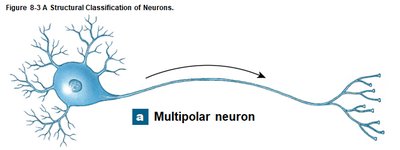
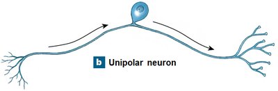
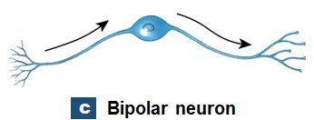
Structural Classification of Neurons
Multipolar Neurons: Many dendrites, one axon. Most common in CNS and all motor neurons.
Unipolar Neurons: Dendrites and axon are continuous, cell body off to one side. Most sensory neurons in PNS.
Bipolar Neurons: One dendrite, one axon, cell body in between. Rare, found in special sense organs.
Functional Classification of Neurons
Sensory (Afferent) Neurons: Transmit sensory information to the CNS.
Motor (Efferent) Neurons: Carry instructions from the CNS to effectors.
Interneurons: Connect sensory and motor neurons within the CNS; involved in higher functions like memory and learning.
Neuroglia (Glial Cells)
Neuroglia support and protect neurons. They are more numerous than neurons and can divide.
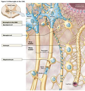
CNS Neuroglia:
Astrocytes: Maintain blood-brain barrier, support neurons
Oligodendrocytes: Produce myelin sheaths in CNS
Microglia: Phagocytic, remove debris and pathogens
Ependymal Cells: Line ventricles, produce and circulate cerebrospinal fluid (CSF)
PNS Neuroglia:
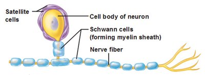
Satellite Cells: Support neuron cell bodies in ganglia
Schwann Cells: Form myelin sheaths around PNS axons
Neural Organization and Physiology
Gray Matter and White Matter
Gray Matter: Contains neuron cell bodies, dendrites, and unmyelinated axons.
White Matter: Contains myelinated axons; myelin gives a glossy white appearance.
Membrane Potential
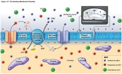
All living cells have a polarized plasma membrane, with a difference in charge across the membrane called the membrane potential. In neurons, the resting membrane potential is typically –70 mV.
Key Factors:
ECF is high in Na+ and Cl–; ICF is high in K+ and negatively charged proteins (Pr–).
Leak channels and gated channels regulate ion movement.
Sodium–potassium exchange pump maintains resting potential by pumping 3 Na+ out and 2 K+ in, using ATP.
Changes in Membrane Potential
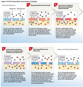
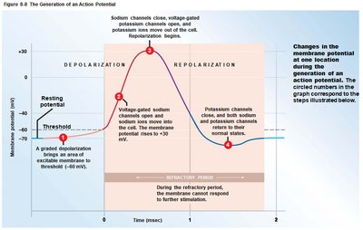
Depolarization: Opening Na+ channels makes the inside less negative (toward 0 mV).
Hyperpolarization: Opening K+ channels makes the inside more negative (further from 0 mV).
Graded Potentials: Local changes that decrease with distance from the stimulus.
Action Potentials: All-or-none electrical impulses that travel the length of the axon if threshold is reached.
Propagation of Action Potentials
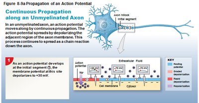
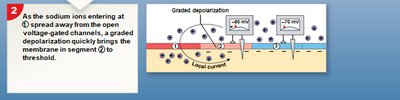
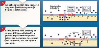
Continuous Propagation: Occurs in unmyelinated axons; action potential moves in a wave along the axon.
Saltatory Propagation: Occurs in myelinated axons; action potential jumps from node to node, increasing speed.
The Synapse
Synapse: Site where a neuron communicates with another cell via neurotransmitters.
Presynaptic Neuron: Releases neurotransmitter into the synaptic cleft.
Postsynaptic Neuron: Receives neurotransmitter via receptors.
Types of Synapses:
Neuromuscular junction (neuron to muscle)
Neuroglandular junction (neuron to gland)
Neurotransmitters
Acetylcholine (ACh): Common neurotransmitter at cholinergic synapses; broken down by acetylcholinesterase (AChE).
Norepinephrine (NE): Released by adrenergic synapses; important in the brain and ANS.
Dopamine, GABA, Serotonin: CNS neurotransmitters with various effects (inhibitory or excitatory).
Excitatory Neurotransmitters: Usually ACh and NE; cause depolarization.
Inhibitory Neurotransmitters: Usually dopamine, GABA, serotonin; cause hyperpolarization.
Organization of the CNS and PNS
Neuron Organization in the PNS
Ganglia: Collections of neuron cell bodies (gray matter) in the PNS, surrounded by satellite cells.
Nerves: Bundles of axons (white matter) in the PNS; can be sensory, motor, or mixed.
Neuron Organization in the CNS
Nuclei: Collections of neuron cell bodies (gray matter) in the CNS.
Tracts: Bundles of axons (white matter) in the CNS; form columns in the spinal cord.
Neural Cortex: Thick layer of gray matter covering parts of the brain surface.
Summary Table: Structural and Functional Organization
Region | Gray Matter | White Matter |
|---|---|---|
CNS | Nuclei, neural cortex | Tracts, columns |
PNS | Ganglia | Nerves |
Additional info: This guide covers the structure and function of the nervous system, including cellular components, membrane physiology, and the organization of neural pathways. It is suitable for introductory college-level anatomy and physiology courses.
