 Back
The Nervous System: Structure, Function, and Clinical Correlates
Study Guide - Smart Notes
 Tailored notes based on your materials, expanded with key definitions, examples, and context.
The Nervous System: Overview
Central and Peripheral Nervous Systems
The nervous system is divided into the Central Nervous System (CNS) and the Peripheral Nervous System (PNS). The CNS consists of the brain and spinal cord, while the PNS includes all neural tissue outside the CNS. The PNS is further subdivided into the somatic and autonomic nervous systems, with the autonomic system divided into sympathetic and parasympathetic divisions.
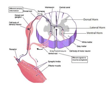
Motor (efferent) signals: Originate in the CNS and travel to effectors (muscles, glands, organs).
Sensory (afferent) signals: Originate in the PNS and are sent to the CNS for processing.
Comparison of Somatic and Autonomic Nervous Systems
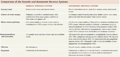
The somatic and autonomic nervous systems differ in their control, effectors, and neurotransmitters.
Somatic Nervous System | Autonomic Nervous System | |
|---|---|---|
Sensory Input | From somatic senses and special senses | Mainly from interoceptors, some from somatic and special senses |
Control of Motor Output | Voluntary (cerebral cortex) | Involuntary (hypothalamus, brainstem, spinal cord) |
Motor Neuron Pathway | One-neuron pathway | Two-neuron pathway (preganglionic and postganglionic) |
Neurotransmitters | Acetylcholine (ACh) | ACh, norepinephrine (NE), epinephrine |
Effectors | Skeletal muscle | Cardiac muscle, smooth muscle, glands |
Responses | Contraction of skeletal muscle | Contraction/relaxation of muscle, secretion of glands |
Neuron Structure and Types
Basic Structure of a Neuron
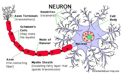
Neurons are the functional units of the nervous system, specialized for communication. They consist of a cell body (soma), dendrites (receive signals), and an axon (transmits signals). The axon may be covered by a myelin sheath, which increases the speed of impulse conduction.
Axon terminals: Release neurotransmitters to communicate with other cells.
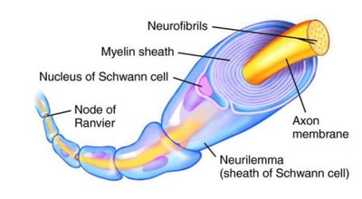
Schwann cells: Produce myelin in the PNS.
Nodes of Ranvier: Gaps in the myelin sheath that facilitate rapid conduction.
Types of Neurons
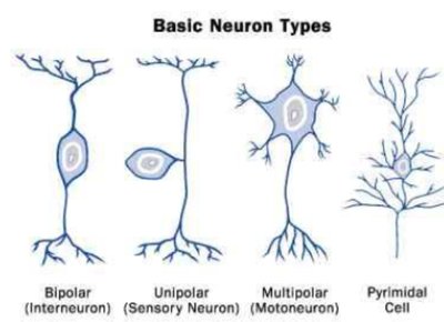
Neurons are classified based on structure and function:
Bipolar neurons: One axon and one dendrite (e.g., retina).
Unipolar neurons: Single process that splits into two branches (sensory neurons).
Multipolar neurons: One axon, multiple dendrites (most common, e.g., motor neurons).
Pyramidal cells: Found in the cerebral cortex.
Neuroglia (Glial Cells)
Types and Functions of Neuroglia
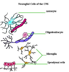
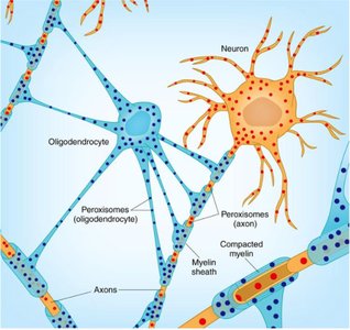
Neuroglia are support cells in the nervous system. They do not conduct impulses but are essential for neuron function and health. They can undergo mitosis, unlike neurons.
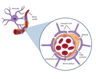
Astrocytes: Maintain electrolyte balance, form part of the blood-brain barrier.
Oligodendrocytes: Produce myelin in the CNS.
Microglia: Phagocytic cells that remove debris and attack invaders.
Ependymal cells: Produce cerebrospinal fluid (CSF).
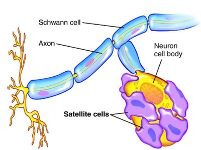
Schwann cells: Produce myelin in the PNS.
Satellite cells: Support ganglia in the PNS.
Synapses and Neurotransmission
Structure and Function of Synapses
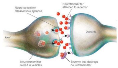
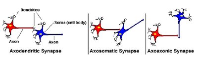
A synapse is the junction between two neurons or between a neuron and an effector cell. It allows for the transmission of electrical or chemical signals. Types of synapses include axodendritic, axosomatic, and axoaxonic.
Neurotransmitters are released from the presynaptic neuron and bind to receptors on the postsynaptic cell.
Neurotransmitter removal occurs via enzyme degradation, diffusion, or reuptake.
Diseases and Disorders of the Nervous System
Multiple Sclerosis (MS)
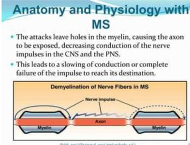
MS is an autoimmune disease where the immune system attacks the myelin sheath, leading to impaired nerve conduction and the formation of plaques in the CNS.
Symptoms include muscle weakness, coordination problems, and sensory disturbances.
Tetanus and Botulism
Tetanus: Caused by Clostridium tetani toxin, which blocks inhibitory neurotransmitter release, resulting in spastic paralysis.
Botulism: Caused by Clostridium botulinum toxin, which inhibits acetylcholine release, resulting in flaccid paralysis.
Spinal Cord Structure and Function
Gray and White Matter
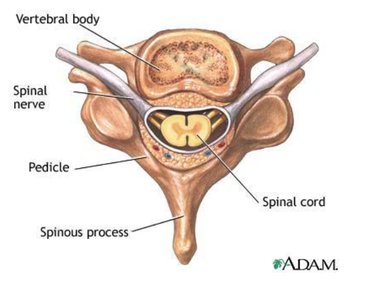
The spinal cord contains gray matter (cell bodies, unmyelinated) and white matter (myelinated axons). The anterior (ventral) horn is motor, and the posterior (dorsal) horn is sensory.
Spinal Cord Termination and Associated Structures
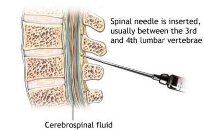
Conus medullaris: End of the solid spinal cord (at L1).
Cauda equina: Bundle of nerve roots below L1.
Filum terminale: Extension of pia mater anchoring the cord to the coccyx.
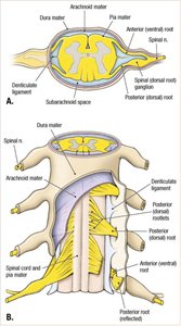
Denticulate ligaments: Extensions of pia mater anchoring the cord laterally.
Clinical Correlates: Shingles and Poliovirus
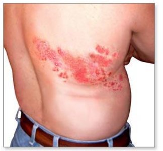
Shingles: Reactivation of Varicella Zoster Virus in dorsal root ganglia, causing painful rash.
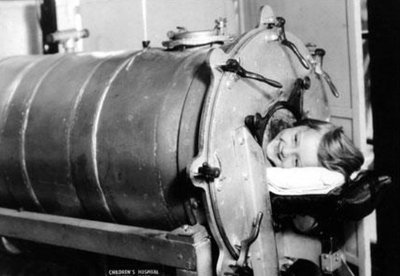
Poliovirus: Attacks anterior horn cells, leading to paralysis and respiratory failure (iron lung may be required).
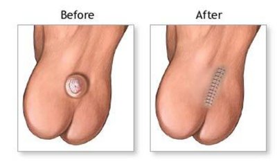
Spina Bifida
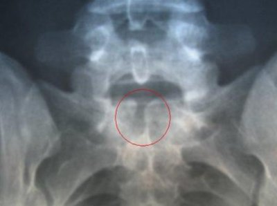
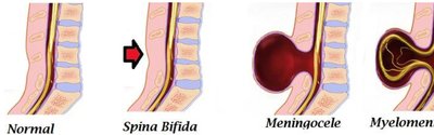
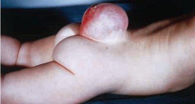
Spina bifida is a neural tube defect where the vertebral column does not close completely. Types include spina bifida occulta, meningocele, and myelomeningocele.
Brain Structure and Function
Major Brain Regions
Cerebrum: Responsible for higher functions such as reasoning, personality, and interpretation.
Cerebellum: Coordinates balance and muscle activity, involved in timekeeping and sensory evaluation.
Corpus callosum: Connects left and right cerebral hemispheres.
Thalamus: Relay center for sensory information.
Hypothalamus: Regulates hormones, appetite, thirst, temperature, and other autonomic functions.
Pituitary gland: Controlled by the hypothalamus, releases hormones affecting many body systems.
Brain stem: Includes midbrain, pons, and medulla oblongata; controls vital reflexes.
Cerebrospinal Fluid (CSF) and Meninges
CSF is produced by the choroid plexus in the ventricles and circulates through the brain and spinal cord, providing protection and nutrient transport. The meninges are three connective tissue layers: pia mater, arachnoid mater, and dura mater.
Pia mater: Innermost, delicate layer.
Arachnoid mater: Middle layer; subarachnoid space contains CSF.
Dura mater: Outermost, tough layer.
Blood-Brain Barrier
The blood-brain barrier is formed by tight junctions between endothelial cells, supported by astrocytes, and protects the brain from harmful substances.
Cranial Nerves
Overview and Functions
There are twelve pairs of cranial nerves, each with specific sensory and/or motor functions, including control of eye movement, facial expression, hearing, taste, and autonomic functions.
Oculomotor: Eye movement, pupil constriction.
Trigeminal: Sensation from face, mastication.
Facial: Facial expression, taste, salivation.
Vestibulocochlear: Hearing and balance.
Vagus: Autonomic control of thoracic and abdominal organs.
Summary Table: Key Neuroanatomical Structures and Functions
Structure | Function |
|---|---|
Cerebrum | Thinking, reasoning, personality |
Cerebellum | Balance, muscle coordination |
Thalamus | Sensory relay |
Hypothalamus | Homeostasis, hormone control |
Medulla oblongata | Vital reflexes (heart rate, breathing) |
Corpus callosum | Connects hemispheres |
Additional info:
Neurotransmitter removal is crucial for synaptic function; SSRIs block serotonin reuptake to treat depression.
Hydrocephalus results from impaired CSF flow, leading to increased intracranial pressure.
Bacterial meningitis is a medical emergency due to the blood-brain barrier limiting antibiotic access.
