 Back
BackThe Peripheral Nervous System and Cranial Nerves: Structure, Function, and Clinical Relevance
Study Guide - Smart Notes
 Tailored notes based on your materials, expanded with key definitions, examples, and context.
The Peripheral Nervous System (PNS)
Overview and Organization
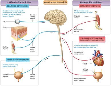
The Peripheral Nervous System (PNS) consists of all neural structures outside the brain and spinal cord. It connects the Central Nervous System (CNS) to limbs and organs, serving as a communication relay between the brain, spinal cord, and the rest of the body.
Somatic Sensory Division: Transmits sensory information from the skin, muscles, and joints to the CNS.
Visceral Sensory Division: Conveys sensory information from internal organs.
Somatic Motor Division: Sends motor commands from the CNS to skeletal muscles.
Visceral Motor Division (Autonomic Nervous System): Regulates involuntary functions by controlling smooth muscle, cardiac muscle, and glands.
Anatomy of Spinal Nerves
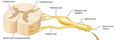
Spinal nerves are mixed nerves carrying both sensory and motor fibers. Each spinal nerve is formed by the union of an anterior (ventral) root (motor fibers) and a posterior (dorsal) root (sensory fibers). The posterior root contains a ganglion housing sensory neuron cell bodies.
Anterior root: Contains motor (efferent) fibers from the CNS to muscles and glands.
Posterior root: Contains sensory (afferent) fibers from receptors to the CNS.
Spinal nerve: Short, mixed nerve formed by the fusion of anterior and posterior roots.
Structure of a Spinal Nerve
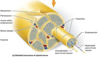
Each spinal nerve is composed of bundles of axons (nerve fibers) organized into fascicles, surrounded by connective tissue layers:
Endoneurium: Surrounds individual axons.
Perineurium: Encloses each fascicle (bundle of axons).
Epineurium: Outermost layer, encasing the entire nerve.
The Cranial Nerves
Overview
There are 12 pairs of cranial nerves, each with specific sensory, motor, or mixed functions. They emerge directly from the brain and brainstem and primarily serve the head and neck regions.
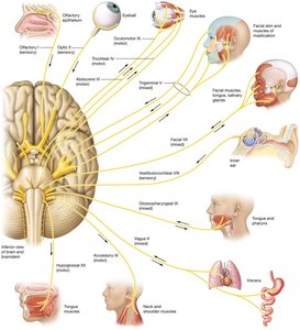
Cranial Nerve I: Olfactory Nerve
The olfactory nerve (I) is responsible for the sense of smell. It contains chemoreceptors that depolarize in response to airborne chemicals, transmitting signals to the primary olfactory cortex in the brain.

Cranial Nerve II: Optic Nerve
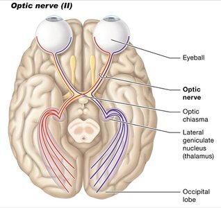
The optic nerve (II) transmits visual information from the retina to the brain. Action potentials are generated when light stimulates photoreceptors in the eye, and these signals are processed by the primary visual cortex.
Cranial Nerves III, IV, VI: Oculomotor, Trochlear, and Abducens Nerves
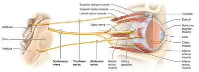
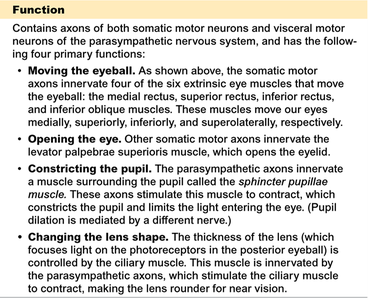
These nerves control most of the eye's movements, pupil constriction, and lens shape. The oculomotor nerve (III) also innervates muscles that open the eyelid and adjust the lens for near vision.
Oculomotor (III): Moves the eye medially, superiorly, inferiorly, and superolaterally; opens the eyelid; constricts the pupil; changes lens shape.
Trochlear (IV): Innervates the superior oblique muscle (moves eye inferiorly and laterally).
Abducens (VI): Innervates the lateral rectus muscle (moves eye laterally).
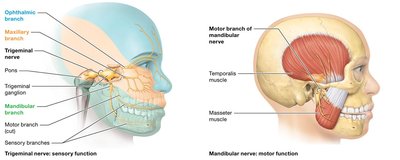
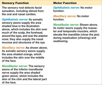
Cranial Nerve V: Trigeminal Nerve
The trigeminal nerve (V) is the main sensory nerve of the face and also controls muscles involved in mastication (chewing). It has three branches: ophthalmic, maxillary, and mandibular.
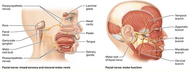
Cranial Nerve VII: Facial Nerve
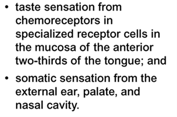
The facial nerve (VII) controls muscles of facial expression and conveys taste sensations from the anterior two-thirds of the tongue. It also provides somatic sensation from the external ear, palate, and nasal cavity.
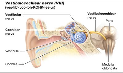
Cranial Nerve VIII: Vestibulocochlear Nerve
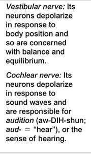
The vestibulocochlear nerve (VIII) is responsible for hearing and balance. It has two components:
Vestibular nerve: Senses body position and equilibrium.
Cochlear nerve: Senses sound waves for hearing.
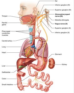
Cranial Nerves IX and X: Glossopharyngeal and Vagus Nerves
The glossopharyngeal nerve (IX) provides taste sensation from the posterior third of the tongue and controls swallowing. The vagus nerve (X) has extensive parasympathetic functions throughout the thoracic and abdominal organs (covered in detail in the autonomic nervous system chapter).
Additional Key Concepts
Sensory Transduction and Receptors
Sensory transduction is the process by which sensory receptors convert external stimuli into electrical signals. Receptors can be classified by location (exteroceptors, interoceptors) or by stimulus type (mechanoreceptors, thermoreceptors, chemoreceptors, photoreceptors, nociceptors).
Encapsulated nerve endings: Surrounded by supportive cells, often rapidly adapting.
Free nerve endings: Lack specialized support, often slowly adapting.
Reflex Arcs and Somatic Reflexes
A reflex arc is a neural pathway that mediates a reflex action. Reflexes can be monosynaptic (single synapse) or polysynaptic (multiple synapses). Major somatic reflexes include the stretch reflex, Golgi tendon reflex, flexion (withdrawal) reflex, and crossed-extension reflex.
Muscle spindles: Detect muscle stretch and initiate the stretch reflex.
Golgi tendon organs: Monitor muscle tension and mediate the tendon reflex to prevent damage.
Clinical Relevance
Understanding the structure and function of the PNS and cranial nerves is essential for diagnosing neurological disorders, localizing lesions, and understanding sensory and motor deficits.
