 Back
BackCh. 13 The Peripheral Nervous System and Reflex Activity: Structure, Function, and Organization
Study Guide - Smart Notes
 Tailored notes based on your materials, expanded with key definitions, examples, and context.
The Peripheral Nervous System (PNS) in the Structural Organization of the Nervous System
Overview of Nervous System Organization
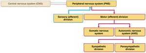
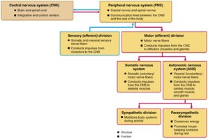
The nervous system is divided into the central nervous system (CNS) and the peripheral nervous system (PNS). The CNS consists of the brain and spinal cord, serving as the main control center. The PNS connects the CNS to the rest of the body and is further subdivided into sensory (afferent) and motor (efferent) divisions.
Sensory (Afferent) Division: Transmits sensory information from receptors to the CNS.
Motor (Efferent) Division: Transmits commands from the CNS to effector organs (muscles and glands).
The motor division is further divided into the somatic nervous system (voluntary control of skeletal muscles) and the autonomic nervous system (ANS) (involuntary control of smooth muscle, cardiac muscle, and glands).
The ANS is subdivided into the sympathetic and parasympathetic divisions.
Sensory Receptors and Their Classification
Types and Functions of Sensory Receptors
Sensory receptors are specialized cells or structures that detect changes in the environment (stimuli) and initiate nerve impulses. Sensation is the awareness of these changes, while perception is the interpretation of their meaning, both occurring in the brain.
Mechanoreceptors: Detect touch, pressure, vibration, and stretch (e.g., in skin, muscles, joints).
Thermoreceptors: Respond to temperature changes (e.g., in skin).
Photoreceptors: Respond to light (e.g., retina of the eye).
Chemoreceptors: Detect chemicals (e.g., smell, taste, blood chemistry).
Nociceptors: Detect pain from damaging stimuli (e.g., extreme heat/cold, pressure, chemicals).
Baroreceptors: Detect pressure changes (e.g., blood pressure in arteries).
Classification by Location
Exteroceptors: Respond to external stimuli (e.g., touch, temperature, pain in skin; special senses).
Interoceptors (Visceroceptors): Respond to internal stimuli (e.g., chemical changes, stretch, temperature in viscera and blood vessels).
Proprioceptors: Detect stretch and position in muscles, tendons, joints, and connective tissues, informing the brain about body movements.
Sensory Processing
Sensation and Perception
Survival depends on the ability to detect (sensation) and interpret (perception) changes in the internal and external environment. Sensory processing involves the conversion of stimuli into electrical signals (graded potentials) that can trigger nerve impulses.
Structure of a Nerve
Connective Tissue Coverings
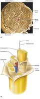
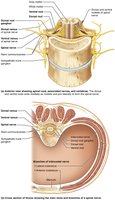
Nerves are organized into bundles of axons, each surrounded by connective tissue layers:
Endoneurium: Surrounds individual axons and their myelin sheaths.
Perineurium: Bundles groups of axons into fascicles.
Epineurium: Encloses all fascicles to form the complete nerve.
Cranial Nerves
Overview and Classification
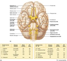
There are 12 pairs of cranial nerves, each associated with the brain. Two pairs attach to the forebrain, while the rest connect to the brainstem. Most cranial nerves are mixed (sensory and motor), but some are purely sensory.
Numbered I–XII from rostral (front) to caudal (back).
Mnemonic for functions (sensory, motor, both): "Some say marry money, but my brother believes (it’s) bad business (to) marry money."
Spinal Nerves
Classification and Distribution
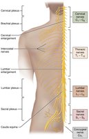
There are 31 pairs of spinal nerves, all mixed nerves named according to their point of origin from the spinal cord. They supply all body parts except the head and part of the neck.
8 pairs of cervical nerves (C1–C8)
12 pairs of thoracic nerves (T1–T12)
5 pairs of lumbar nerves (L1–L5)
5 pairs of sacral nerves (S1–S5)
1 pair of coccygeal nerves (Co1)
Innervation of Specific Body Regions
Nerve Plexuses
All ventral rami (except thoracic) form interlacing networks called nerve plexuses in the cervical, brachial, lumbar, and sacral regions. Plexuses allow fibers from different spinal nerves to combine and redistribute, ensuring that each limb muscle is innervated by more than one spinal nerve. This redundancy prevents complete paralysis if one nerve is damaged.
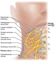
Cervical Plexus and the Neck
The cervical plexus is formed by the first four ventral rami (C1–C4). It provides cutaneous nerves to the skin of the neck, ear, back of the head, and shoulders, and motor branches to neck muscles. The phrenic nerve is the major motor and sensory nerve of the diaphragm, essential for breathing.
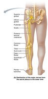
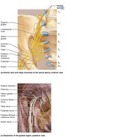
Sacral Plexus and Lower Limb
The sacral plexus arises from the lower lumbar and sacral spinal nerves. It serves the buttock, lower limb, pelvic structures, and perineum. The sciatic nerve is the longest and thickest nerve in the body, innervating the hamstrings, adductor magnus, and most muscles in the leg and foot. It is composed of the tibial and common fibular nerves.

Reflex Arcs and Reflex Activity
Components of a Reflex Arc
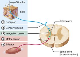
A reflex arc is the neural pathway that mediates a reflex. It consists of five basic components:
Receptor: Site of stimulus action.
Sensory neuron: Transmits afferent impulses to the CNS.
Integration center: Synapse(s) within the CNS (monosynaptic or polysynaptic).
Motor neuron: Conducts efferent impulses from the integration center to an effector.
Effector: Muscle or gland that responds to the motor neuron impulse.
Types of Reflexes
Somatic reflexes: Activate skeletal muscle.
Autonomic (visceral) reflexes: Activate smooth muscle, cardiac muscle, or glands.
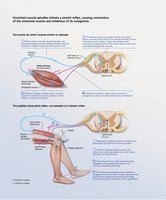
Stretch Reflex
The stretch reflex is initiated by muscle spindles and causes contraction of the stretched muscle and inhibition of its antagonist. This helps maintain muscle tone and posture.
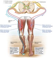
Crossed-Extensor Reflex
The crossed-extensor reflex is a complex spinal reflex that helps maintain balance. When one limb withdraws from a painful stimulus, the opposite limb extends to support the body.
Additional info: The organization and function of the PNS, including cranial and spinal nerves, nerve plexuses, and reflex arcs, are essential for understanding how the nervous system controls and coordinates body activities. Damage to these structures can result in sensory or motor deficits, emphasizing the importance of redundancy and integration in neural pathways.
