 Back
The Peripheral Nervous System: Structure, Function, and Integration
Study Guide - Smart Notes
 Tailored notes based on your materials, expanded with key definitions, examples, and context.
The Peripheral Nervous System (PNS)
Overview and Organization
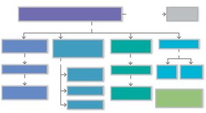
The Peripheral Nervous System (PNS) connects the central nervous system (CNS) to sensory receptors, muscles, and glands throughout the body. It is responsible for gathering sensory input and executing motor output, thus facilitating communication between the body and the CNS.
Sensory (afferent) division: Transmits sensory information from receptors to the CNS.
Motor (efferent) division: Transmits motor commands from the CNS to effectors (muscles and glands).
The motor division is further divided into the somatic nervous system (voluntary control of skeletal muscles) and the autonomic nervous system (ANS) (involuntary control of smooth muscle, cardiac muscle, and glands).
The ANS is subdivided into the sympathetic and parasympathetic divisions.
Sensory Receptors and Sensory Integration
Sensory Receptors: Definition and Function
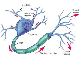
Sensory receptors are specialized structures that respond to changes in the environment (stimuli). When activated by an adequate stimulus, they generate graded potentials that can trigger nerve impulses along afferent fibers toward the CNS.
Sensation: Awareness of the stimulus.
Perception: Interpretation of the meaning of the stimulus.
Receptive Fields
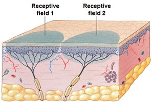
Each sensory receptor monitors a specific area known as its receptive field. The stimulus must be applied within this field to activate the receptor.
Processing at the Receptor Level
Stimulus energy must match the receptor's specificity.
Stimulus must be applied within the receptor's receptive field.
Transduction: Conversion of stimulus energy into a graded potential.
Types of graded potentials:
Generator potential: Occurs when the receptor region is part of a sensory neuron (general senses).
Receptor potential: Occurs when the receptor is a separate cell (special senses).
Adaptation of Sensory Receptors
Adaptation is a decrease in receptor sensitivity in the presence of a constant stimulus. Receptors can be classified as:
Phasic receptors: Fast-adapting (e.g., pressure, touch, smell).
Tonic receptors: Slow-adapting or non-adapting (e.g., pain, proprioceptors).
Classification of Sensory Receptors
Sensory receptors can be classified by:
Stimulus type: Mechanoreceptors, thermoreceptors, photoreceptors, chemoreceptors, nociceptors.
Location: Exteroceptors, interoceptors, proprioceptors.
Structural complexity: Simple (general senses) or complex (special senses).
General Sensory Receptors: Structure and Function
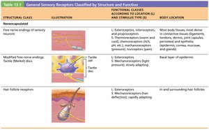
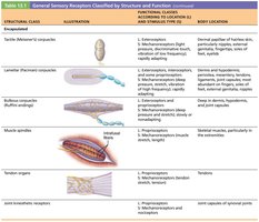
General sensory receptors are classified by their structure and function. The following tables summarize the main types:
Structural Class | Illustration | Functional Classes | Body Location |
|---|---|---|---|
Free nerve endings of sensory neurons |
| Exteroceptors, interoceptors, proprioceptors; thermoreceptors, chemoreceptors, mechanoreceptors, nociceptors | Most body tissues; connective tissues, epithelia |
Modified free nerve endings (Merkel discs) |
| Exteroceptors; mechanoreceptors (light pressure, slowly adapting) | Basal layer of epidermis |
Hair follicle receptors | Exteroceptors; mechanoreceptors (hair deflection, rapidly adapting) | In and surrounding hair follicles |

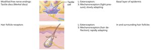
Encapsulated dendritic endings include Meissner's corpuscles, Pacinian corpuscles, Ruffini endings, muscle spindles, Golgi tendon organs, and joint kinesthetic receptors.
Nerve Fibers and Repair
Structure of a Nerve
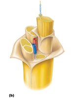
Nerves are cordlike organs consisting of parallel bundles of peripheral axons (fibers) enclosed by connective tissue wrappings: endoneurium, perineurium, and epineurium.
Regeneration of Nerve Fibers
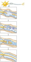
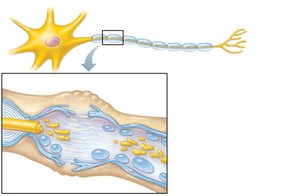
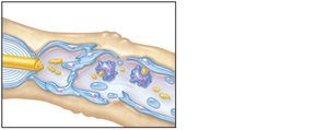
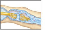
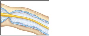
Peripheral nerves can regenerate if the cell body remains intact. The process involves several steps:
Axon fragments and myelin sheaths degenerate distal to injury (Wallerian degeneration).
Schwann cells and macrophages clean out the dead axon distal to the injury.
Axon filaments grow through a regeneration tube formed by Schwann cells.
The axon regenerates and a new myelin sheath forms.
Processing of Sensory Information
Levels of Neural Integration
Receptor level: Sensory receptors detect stimuli.
Circuit level: Ascending pathways deliver impulses to the appropriate region of the cerebral cortex.
Perceptual level: Neuronal circuits in the cerebral cortex interpret sensory input.
Processing at the Circuit Level
Three-neuron pathways conduct sensory impulses upward:
First-order neurons (PNS) → Second-order neurons (spinal cord/CNS) → Thalamus or cerebellum
Third-order neurons → Somatosensory cortex (perceptual level)
Processing at the Perceptual Level
Sensory input is interpreted in the cerebral cortex. Each sensory fiber corresponds to a "labeled line" that tells the brain the type and location of the stimulus.
Perceptual detection: Detecting a stimulus.
Magnitude estimation: Determining stimulus intensity.
Spatial discrimination: Identifying the site of stimulation.
Feature abstraction: Identifying complex aspects of a stimulus.
Quality discrimination: Differentiating submodalities (e.g., sweet vs. sour).
Pattern recognition: Recognizing familiar patterns (e.g., melody).
Perception of Pain and Referred Pain
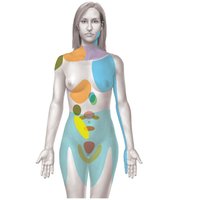
Pain warns of actual or impending tissue damage. Referred pain occurs because somatic and autonomic sensory signals are carried along parallel nerve tracts, causing pain to be perceived at a site other than the actual origin.
Motor Endings and Motor Activity
Levels of Motor Control
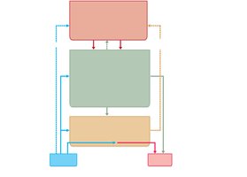
Motor control is organized into three hierarchical levels:
Segmental Level (lowest): Spinal cord circuits control reflexes and central pattern generators (CPGs) for repetitive movements.
Projection Level (middle): Motor cortex and brain stem nuclei convey instructions to spinal cord motor neurons.
Precommand Level (highest): Cerebellum and basal nuclei regulate and coordinate movements.
Reflexes and Reflex Arcs
Definition and Function
A reflex is a fast, predictable, automatic response to changes in the environment. Reflexes help maintain homeostasis and are integrated in the gray matter of the spinal cord.
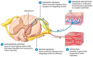
General Components of a Reflex Arc
Receptor: Detects the stimulus.
Sensory neuron: Transmits afferent impulses to the CNS.
Integration center: One or more synapses in the CNS.
Motor neuron: Conducts efferent impulses to an effector.
Effector: Muscle or gland that responds to the impulse.
Types of Reflexes
Stretch reflex: Maintains muscle length by causing contraction of the stretched muscle and inhibition of its antagonist.
Tendon reflex: Controls muscle tension by causing muscle relaxation when tension is excessive.
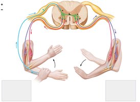
Flexor reflex: Withdrawal of a limb from a painful stimulus.
Crossed extensor reflex: Opposite limb extends to maintain balance during a flexor reflex.
Additional info: This guide covers the structure and function of the peripheral nervous system, sensory receptors, neural integration, nerve fiber repair, and reflexes, providing a comprehensive overview for ANP college students.
