 Back
BackThe Peripheral Nervous System: Structure, Function, and Clinical Relevance
Study Guide - Smart Notes
 Tailored notes based on your materials, expanded with key definitions, examples, and context.
Chapter 13: The Peripheral Nervous System
Overview of the Peripheral Nervous System (PNS)
The Peripheral Nervous System (PNS) serves as the communication link between the Central Nervous System (CNS) and the rest of the body. It detects sensory stimuli and delivers this information to the CNS as sensory input. The CNS processes this input and sends motor output through the PNS to muscle cells and glands.
Sensory (Afferent) Division: Transmits sensory information from receptors to the CNS.
Motor (Efferent) Division: Transmits motor commands from the CNS to effectors (muscles and glands).
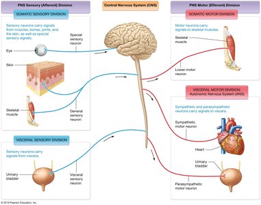
Peripheral Nerves and Associated Ganglia
Peripheral nerves are bundles of axons that connect the CNS to the body. They are classified as either spinal nerves (originating from the spinal cord) or cranial nerves (originating from the brain).
Spinal Nerves: Originate from the spinal cord and innervate structures below the head and neck.
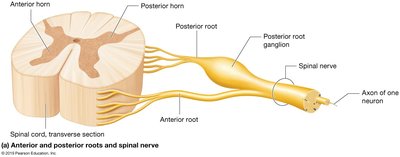
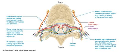
Each spinal nerve is formed by the union of two roots:
Anterior root: Contains motor neurons from the anterior horn.
Posterior root: Contains sensory neurons from the posterior horn.
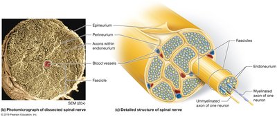
Spinal nerves are organized into fascicles, each surrounded by connective tissue:
Epineurium: Outermost layer, holds motor and sensory axons together.
Perineurium: Surrounds each fascicle (bundle of axons).
Endoneurium: Surrounds each individual axon within a fascicle.
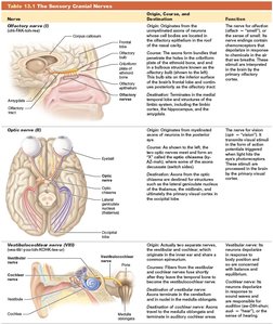
Cranial Nerves
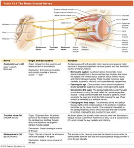
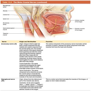
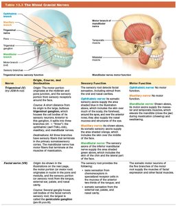
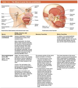
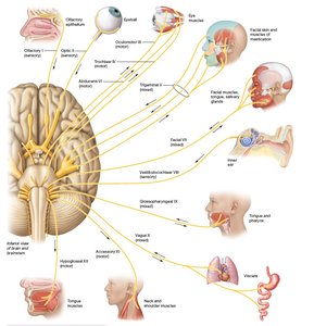
Cranial nerves attach to the brain and primarily innervate structures in the head and neck. They can be classified as sensory, motor, or mixed nerves.
Sensory Cranial Nerves: Olfactory (I), Optic (II), Vestibulocochlear (VIII)
Motor Cranial Nerves: Oculomotor (III), Trochlear (IV), Abducens (VI), Accessory (XI), Hypoglossal (XII)
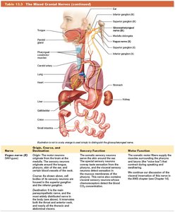
Mixed Cranial Nerves: Trigeminal (V), Facial (VII), Glossopharyngeal (IX), Vagus (X)
Clinical Application: Bell's Palsy
Bell's palsy is a condition where the facial nerve (VII) is impaired, leading to weakness or paralysis of facial muscles on one side. This can affect blinking, closing the eye, and facial expressions.

Spinal Nerves and Plexuses
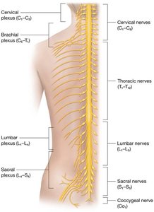
There are 31 pairs of spinal nerves, each formed by the fusion of anterior and posterior roots. Each spinal nerve divides into:
Posterior ramus: Innervates the posterior body.
Anterior ramus: Innervates the anterior body and limbs.
Cervical Plexus
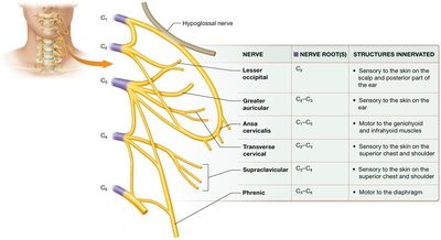
The cervical plexus (C1–C5) innervates the skin and muscles of the neck, head, chest, and shoulders. The phrenic nerve (C3–C5) is a major branch that innervates the diaphragm, essential for breathing.
Brachial Plexus
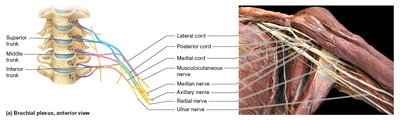
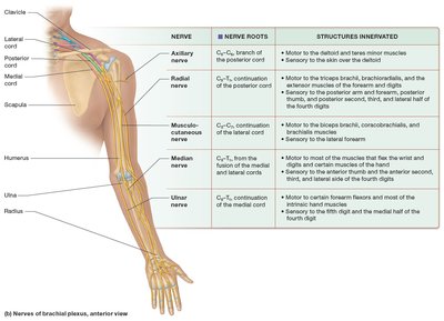
The brachial plexus (C5–T1) provides motor and sensory innervation to the upper limbs. Major nerves include:
Axillary nerve: Deltoid and teres minor muscles, skin over deltoid.
Radial nerve: Triceps brachii, extensor muscles of forearm, skin over posterior thumb and fingers.
Musculocutaneous nerve: Biceps brachii, skin of lateral arm.
Median nerve: Wrist and digital flexors, some hand muscles, skin over anterior thumb and fingers.
Ulnar nerve: Flexor muscles in forearm, most intrinsic hand muscles, skin of 5th digit and medial 4th digit.
Lumbar Plexus
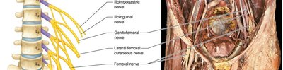
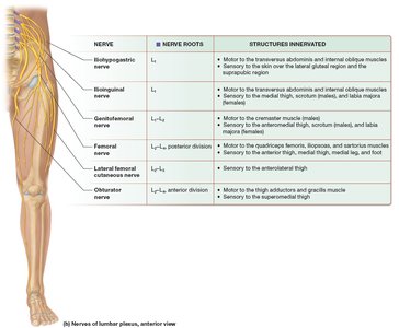
The lumbar plexus (L1–L5) innervates the pelvic region and lower extremity. Major nerves include:
Obturator nerve: Adductor muscles of thigh, hip joint, skin over medial thigh.
Femoral nerve: Anterior thigh muscles, skin over anterior thigh and leg, knee joint.
Sacral Plexus
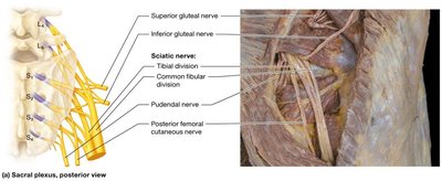
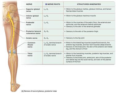
The sacral plexus (L4–S4) innervates the pelvis, gluteal region, and much of the lower limb. Major nerves include:
Sciatic nerve: Longest nerve in the body; divides into tibial and common fibular nerves.
Tibial nerve: Hamstring muscles, knee and ankle joints, plantar flexor muscles.
Common fibular nerve: Lateral leg, dorsum of foot, ankle dorsiflexors.
Classification of Sensory Receptors
Sensory receptors are specialized to detect specific types of stimuli and can be classified by location or stimulus type:
Exteroceptors: Detect external stimuli (e.g., touch, temperature).
Interoceptors: Detect internal stimuli (e.g., blood pressure, pH).
Mechanoreceptors: Respond to mechanical deformation (e.g., pressure, vibration).
Thermoreceptors: Respond to temperature changes.
Chemoreceptors: Respond to chemical changes.
Photoreceptors: Respond to light (found only in the eye).
Nociceptors: Respond to noxious or painful stimuli.
Thermoreceptors are further divided into "cold" (10–40°C) and "hot" (32–48°C) receptors. Temperatures outside these ranges are detected by nociceptors, which is why extreme temperatures are perceived as pain.
Dermatomes and Referred Pain
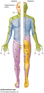
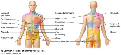
Dermatomes are regions of skin supplied by sensory fibers from a single spinal nerve. Dermatome maps are used clinically to assess sensory pathway integrity. Referred pain occurs when pain from an internal organ is perceived as originating from a dermatome, due to shared neural pathways.
Reflex Arcs: Integration of Sensory and Motor Functions
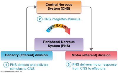
Reflexes are automatic, programmed responses to stimuli, typically protective in nature. They occur via a reflex arc, which involves:
Detection of a stimulus by the PNS and delivery to the CNS.
Integration of the stimulus by the CNS.
Delivery of a motor response by the PNS to effectors.
Reflexes can be classified by:
Number of synapses: Monosynaptic (one synapse) or polysynaptic (multiple synapses).
Type of organ: Somatic (skeletal muscle) or visceral (smooth/cardiac muscle or glands).
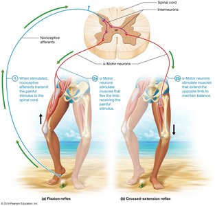
Examples include the flexion reflex and crossed-extension reflex, which protect the body from harm.
Clinical Application: Babinski Sign
The Babinski sign is a clinical test for upper motor neuron disorders. Stroking the bottom of the foot should cause toe flexion in healthy adults (plantar reflex). Extension of the big toe and fanning of other toes is abnormal in adults but normal in infants up to 18 months.
