 Back
BackThe Peripheral Nervous System: Structure, Function, and Integration
Study Guide - Smart Notes
 Tailored notes based on your materials, expanded with key definitions, examples, and context.
Overview of the Peripheral Nervous System (PNS)
Introduction to the PNS
The Peripheral Nervous System (PNS) serves as the communication network linking the Central Nervous System (CNS) to the rest of the body and the external environment. It is responsible for detecting sensory stimuli and transmitting this information to the CNS, which processes the input and sends motor commands back through the PNS to muscles and glands.
Sensory Input: The PNS detects changes in the environment and relays this information to the CNS.
Motor Output: The CNS processes sensory input and sends out motor commands via the PNS to effectors such as muscles and glands.
Divisions of the PNS
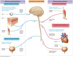
The PNS is functionally divided into sensory and motor divisions, each with further subdivisions:
Sensory (Afferent) Division:
Somatic Sensory Division: Detects stimuli from the skin, muscles, and special sense organs.
Visceral Sensory Division: Relays internal information from organs in the thoracic and abdominopelvic cavities.
Motor (Efferent) Division:
Somatic Motor Division: Controls voluntary movements by innervating skeletal muscles.
Visceral Motor Division (Autonomic Nervous System, ANS): Regulates involuntary functions by innervating cardiac muscle, smooth muscle, and glands. The ANS is further divided into:
Sympathetic Nervous System: Prepares the body for 'fight or flight' responses.
Parasympathetic Nervous System: Promotes 'rest and digest' activities and maintains homeostasis at rest.
Peripheral Nerves and Associated Ganglia
Structure of Peripheral Nerves
Peripheral nerves are the main organs of the PNS, consisting of bundles of axons bound together by connective tissue. They innervate most structures in the body and can be classified as:
Mixed Nerves: Contain both sensory and motor neurons.
Sensory Nerves: Contain only sensory neurons.
Motor Nerves: Contain mostly motor neurons.
Spinal Nerves
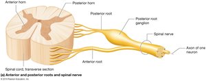
Spinal nerves originate from the spinal cord and innervate structures below the head and neck. Each spinal nerve is formed by the fusion of an anterior (motor) root and a posterior (sensory) root, with the posterior root ganglion housing the cell bodies of sensory neurons. All 31 pairs of spinal nerves are mixed nerves.
Connective Tissue Organization
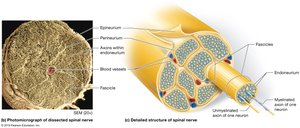
Peripheral nerves are organized into fascicles, each surrounded by connective tissue layers:
Epineurium: Outermost layer, enclosing the entire nerve.
Perineurium: Surrounds each fascicle (bundle of axons).
Endoneurium: Surrounds individual axons within a fascicle.
Cranial Nerves
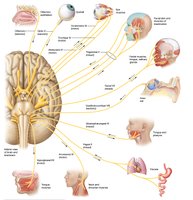
Cranial nerves attach to the brain and primarily innervate structures in the head and neck. Unlike spinal nerves, they are not formed by the fusion of sensory and motor roots and may be purely sensory, mixed, or mostly motor nerves.
Cranial Nerves: Classification and Function
Sensory Cranial Nerves
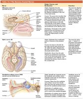
Three cranial nerves contain only sensory axons:
Olfactory (I): Sense of smell
Optic (II): Vision
Vestibulocochlear (VIII): Hearing and balance
Motor Cranial Nerves
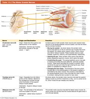
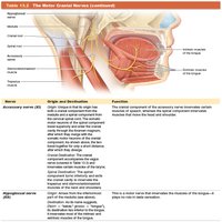
Five cranial nerves are primarily motor, with some proprioceptive sensory axons:
Oculomotor (III): Eye movement, pupil constriction
Trochlear (IV): Eye movement (superior oblique muscle)
Abducens (VI): Eye movement (lateral rectus muscle)
Accessory (XI): Movement of head and shoulders
Hypoglossal (XII): Tongue movement
Mixed Cranial Nerves
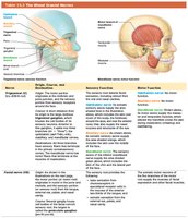
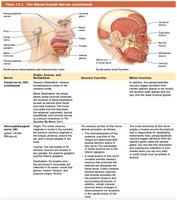
Four cranial nerves contain both sensory and motor axons:
Trigeminal (V): Sensation from face, motor to muscles of mastication
Facial (VII): Taste, facial expression muscles
Glossopharyngeal (IX): Taste, swallowing, salivation
Vagus (X): Sensory and motor to thoracic and abdominal organs
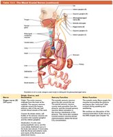
Overview of Cranial Nerves
Spinal Nerves and Plexuses
Structure and Branches of Spinal Nerves
Each spinal nerve divides into two mixed nerves:
Posterior Ramus: Innervates the posterior side of the body.
Anterior Ramus: Innervates the anterior side of the body and limbs.
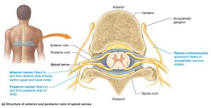
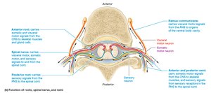
Spinal Nerve Distribution
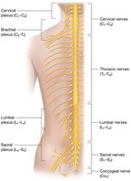
There are 31 pairs of spinal nerves:
8 pairs of cervical nerves
12 pairs of thoracic nerves
5 pairs of lumbar nerves
5 pairs of sacral nerves
1 pair of coccygeal nerves
The anterior rami of cervical, lumbar, and sacral nerves form complex networks called nerve plexuses.
Brachial Plexus
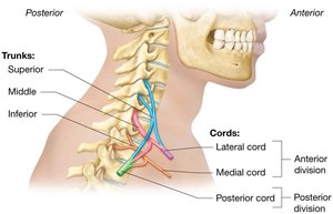
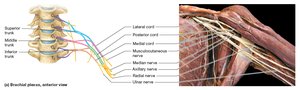
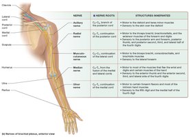
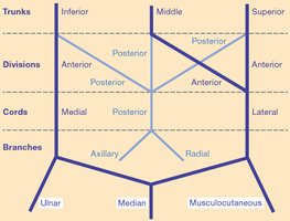
The brachial plexus provides motor and sensory innervation to the upper limbs. It is formed by the anterior rami of C5–T1 and is organized into trunks, divisions, cords, and branches.
Sacral Plexus
The sacral plexus is formed from the anterior rami of L4–S4 and innervates the pelvis, gluteal region, and lower extremity. The sciatic nerve is the largest branch, innervating the posterior thigh and dividing into the tibial and common fibular nerves.
Cutaneous and Motor Distribution
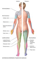
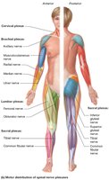
Spinal nerve branches have specific cutaneous (sensory) and motor distributions, which can be mapped on the body surface and muscle groups.
Sensory Reception and Sensory Receptors
Classification of Sensory Receptors
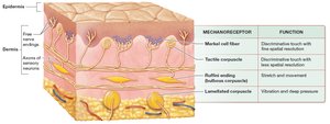
Sensory receptors are specialized to detect specific types of stimuli and can be classified by structure, location, and stimulus type:
Structure: Encapsulated (with supportive cells) or free nerve endings (without supportive cells).
Location: Exteroceptors (external stimuli), Interoceptors (internal stimuli).
Stimulus Type:
Mechanoreceptors: Respond to mechanical deformation (touch, pressure, vibration).
Thermoreceptors: Detect temperature changes.
Chemoreceptors: Respond to chemical changes.
Photoreceptors: Detect light (in the eye).
Nociceptors: Detect pain (noxious stimuli).
Somatic Sensory Neurons
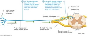
First-order somatic sensory neurons are pseudounipolar, with a peripheral process (from receptor to cell body) and a central process (from cell body to CNS). The cell bodies are located in the dorsal root ganglia (spinal nerves) or cranial nerve ganglia.
Motor Output and Control of Movement
From CNS to PNS: Motor Output
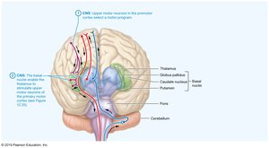
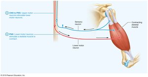
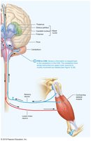
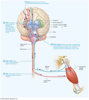
Motor commands originate in the CNS and are transmitted to skeletal muscles via upper and lower motor neurons:
Upper Motor Neurons: Located in the primary motor cortex; initiate voluntary movement but do not directly contact muscle fibers.
Lower Motor Neurons: Located in the anterior horn of the spinal cord or brainstem; directly innervate skeletal muscle fibers and release acetylcholine to trigger contraction.
Reflex Arcs: Integration of Sensory and Motor Functions
Reflex Arcs
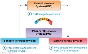
Reflexes are automatic, programmed responses to stimuli that occur via reflex arcs, typically as protective negative feedback loops. Reflexes begin with a sensory stimulus and end with a rapid motor response, with neural integration occurring in the spinal cord or brainstem.
Stretch Receptors in Skeletal Muscle
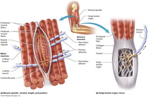
Mechanoreceptors in muscles and tendons monitor muscle length and tension:
Muscle Spindles: Detect changes in muscle length and trigger stretch reflexes.
Golgi Tendon Organs: Monitor muscle tension and trigger relaxation to prevent damage.
Types of Reflexes
Reflexes are classified by the number of synapses (monosynaptic or polysynaptic) and the type of organ involved (somatic or visceral):
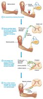
Simple Stretch Reflex: Example: Patellar (knee-jerk) reflex; helps maintain optimal muscle length.
Golgi Tendon Reflex: Prevents excessive muscle tension by causing muscle relaxation.
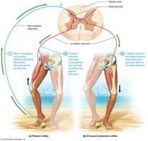
Flexion (Withdrawal) Reflex: Withdraws a limb from a painful stimulus.
Crossed-Extension Reflex: Maintains balance by extending the opposite limb during withdrawal.
Cranial Nerve Reflexes: Involve cranial nerves, e.g., gag reflex and corneal blink reflex.
Additional info: This guide covers the structure and function of the peripheral nervous system, including cranial and spinal nerves, sensory and motor pathways, and reflex integration, as outlined in a typical Anatomy & Physiology curriculum.
