 Back
BackThe Reproductive System: Structure, Function, and Regulation
Study Guide - Smart Notes
 Tailored notes based on your materials, expanded with key definitions, examples, and context.
Chapter 26: The Reproductive System
Introduction to the Male and Female Reproductive Systems
The reproductive systems in males and females are responsible for producing gametes, secreting sex hormones, and supporting the development of offspring. The primary sex organs, or gonads, are the testes in males and the ovaries in females. These organs produce gametes (sperm and ova) and secrete hormones such as testosterone and estrogens. Accessory reproductive organs aid in the transport, nourishment, and protection of gametes and offspring.
Overview of Meiosis
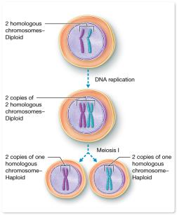
Meiosis is a specialized type of cell division that reduces the chromosome number by half, producing haploid gametes. This process ensures genetic diversity and the correct chromosome number in offspring after fertilization.
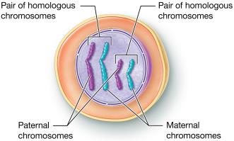
Diploid (2n) cells have 46 chromosomes (23 pairs), with one set from each parent.
Homologous chromosomes carry the same genes but may have different alleles.
Fertilization restores the diploid number by combining haploid sperm and ova to form a zygote.
Concept Boost: Diploid and Haploid Cells
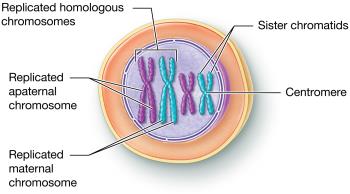
Meiosis reduces the chromosome number from diploid to haploid. After DNA replication, each chromosome consists of two sister chromatids. Meiosis I separates homologous chromosomes, resulting in haploid cells with duplicated chromosomes.
Stages of Meiosis
Meiosis I (Reduction Division): Homologous chromosomes separate, producing haploid cells.
Meiosis II: Sister chromatids separate, resulting in four genetically unique haploid cells.
Comparing Mitosis and Meiosis
Mitosis: Produces two genetically identical diploid somatic cells for growth and repair.
Meiosis: Produces up to four genetically unique haploid gametes for sexual reproduction.
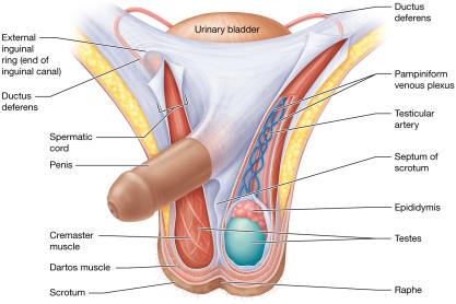
Anatomy of the Male Reproductive System
Internal and External Structures
The male reproductive system includes the testes, duct system, accessory glands, penis, and scrotum. Sperm are produced in the testes and travel through the duct system, where fluids from accessory glands are added to form semen.

Testes
Located in the scrotum, divided into lobules containing seminiferous tubules.
Spermatogenic cells produce sperm; sustentacular cells support development.
Interstitial cells produce testosterone.
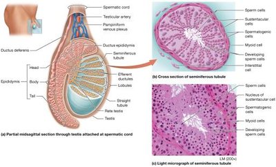
Duct System
Epididymis: Site of sperm maturation and storage.
Ductus (Vas) Deferens: Transports sperm during ejaculation.
Ejaculatory Duct: Joins ductus deferens and seminal vesicle, passes through prostate.
Urethra: Transports urine and semen through prostatic, membranous, and spongy regions.
Penis
Copulatory organ composed of erectile tissue (corpora cavernosa and corpus spongiosum).
Delivers sperm into the female reproductive tract.

Accessory Sex Glands
Seminal Vesicles: Secrete alkaline fluid with fructose, prostaglandins, and coagulating proteins.
Prostate Gland: Secretes milky fluid with citrate, PSA, and antimicrobial chemicals.
Bulbourethral Glands: Secrete alkaline mucus to neutralize urine and lubricate urethra.
Semen
Mixture of sperm and glandular fluids; alkaline pH supports sperm motility and capacitation.
Contains antimicrobial chemicals and prostaglandins.
Support Structures: Scrotum and Spermatic Cord
Scrotum: Sac that houses the testes and regulates temperature for sperm development.
Spermatic Cord: Contains ductus deferens, blood vessels, nerves, and muscles.
Functions of the Male Reproductive Structures
Structure | Function |
|---|---|
Testis | Produces sperm cells, testosterone, and inhibin |
Epididymis | Promotes sperm maturation, stores sperm |
Ductus deferens | Stores and moves sperm |
Ejaculatory duct | Transports sperm to urethra |
Urethra | Transports semen out of penis |
Seminal vesicle | Secretes alkaline fluid with nutrients and enzymes |
Prostate gland | Secretes fluid with nutrients and anticoagulant properties |
Bulbourethral gland | Secretes mucus for lubrication and neutralization |
Scrotum | Protects and regulates temperature of testes |
Penis | Deposits semen in vagina |
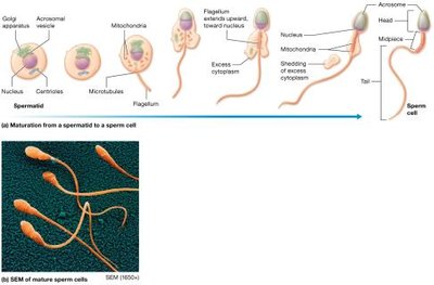
Spermatogenesis and Spermiogenesis
Spermatogenesis
Spermatogenesis is the process of sperm cell development, beginning at puberty and continuing throughout life. It occurs in the seminiferous tubules and involves mitosis, meiosis, and maturation.
Spermatogonia (stem cells) divide by mitosis.
Primary spermatocytes undergo meiosis I to form secondary spermatocytes.
Secondary spermatocytes undergo meiosis II to form spermatids.
Spermiogenesis
Spermiogenesis is the maturation of spermatids into sperm cells, involving the formation of the acrosome, flagellum, and condensation of the nucleus.
Sustentacular (Nurse) Cells
Form the blood-testis barrier, support and nourish developing sperm, and secrete androgen-binding protein and inhibin.
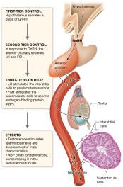
Hormonal Control of Male Reproduction
The hypothalamic-pituitary-gonadal (HPG) axis regulates spermatogenesis and testosterone production through a multi-tiered feedback loop.
GnRH from the hypothalamus stimulates the anterior pituitary to release LH and FSH.
LH stimulates interstitial cells to produce testosterone.
FSH stimulates sustentacular cells to secrete ABP and inhibin.
Testosterone and inhibin provide negative feedback to regulate hormone levels.
Anatomy of the Female Reproductive System
Internal and External Structures
The female reproductive system includes the ovaries, uterine tubes, uterus, vagina, and external genitalia (vulva). These structures support oogenesis, fertilization, and development of the conceptus.
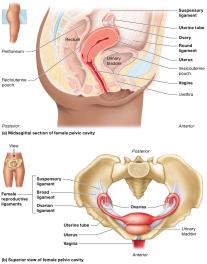
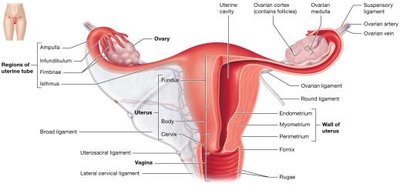
Ovaries
Produce oocytes and secrete estrogens, progesterone, inhibin, and relaxin.
Oogenesis occurs in the ovarian cortex within follicles.
Uterine Tubes (Fallopian Tubes)
Transport oocytes from the ovary to the uterus; site of fertilization.
Fimbriae help capture the oocyte during ovulation.
Uterus
Site of implantation, fetal development, and menstruation.
Composed of perimetrium, myometrium, and endometrium.
Vagina
Receives the penis during intercourse, serves as the birth canal, and allows menstrual flow.
Acidic environment protects against infection.
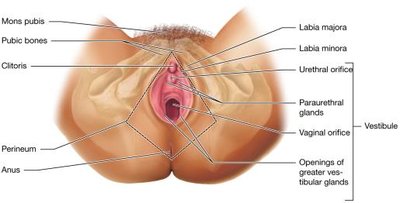
Female External Genitalia (Vulva)
Includes mons pubis, labia majora, labia minora, clitoris, vestibule, and glands for lubrication.
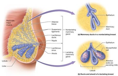
Mammary Glands
Modified sweat glands that produce milk for newborns.
Composed of lobes, lobules, alveoli, and ducts.
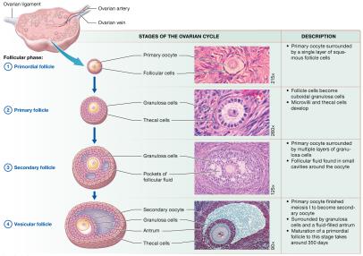
Oogenesis and the Ovarian Cycle
Oogenesis
Oogenesis is the process of ovum development, beginning before birth and continuing until menopause. Primary oocytes are arrested in prophase I until puberty, when the ovarian cycle resumes their development.
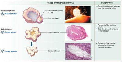
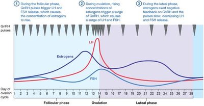
Ovarian Cycle
Follicular Phase: Follicle growth and maturation (primordial, primary, secondary, vesicular follicles).
Ovulation: Release of the secondary oocyte from the ovary.
Luteal Phase: Formation of the corpus luteum, which secretes progesterone and estrogen.
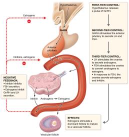
Hormonal Control of the Ovarian Cycle
GnRH stimulates FSH and LH release from the anterior pituitary.
FSH stimulates follicle development; LH triggers ovulation and corpus luteum formation.
Estrogens and inhibin provide feedback to regulate hormone levels.
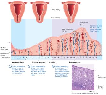
The Uterine (Menstrual) Cycle
The uterine cycle describes cyclic changes in the endometrium in response to ovarian hormones.
Menstrual Phase (Days 1–5): Shedding of the stratum functionalis.
Proliferative Phase (Days 6–14): Regeneration of the endometrium; ends with ovulation.
Secretory Phase (Days 15–28): Endometrium prepares for implantation; if no fertilization, menstruation begins.
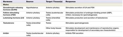
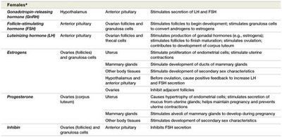
Major Reproductive Hormones in Males and Females
Hormone | Source | Target Tissue(s) | Response |
|---|---|---|---|
GnRH | Hypothalamus | Anterior pituitary | Stimulates secretion of LH and FSH |
FSH | Anterior pituitary | Testes/Ovaries | Stimulates spermatogenesis/follicle development |
LH | Anterior pituitary | Testes/Ovaries | Stimulates testosterone/estrogen and ovulation |
Testosterone | Testes | Various | Development of male characteristics |
Estrogens | Ovaries | Various | Development of female characteristics |
Progesterone | Ovaries (corpus luteum) | Uterus, mammary glands | Prepares endometrium, maintains pregnancy |
Inhibin | Testes/Ovaries | Anterior pituitary | Inhibits FSH secretion |
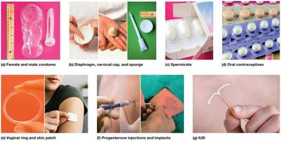
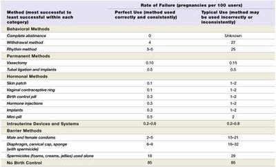
Methods of Birth Control
Behavioral Methods: Abstinence, rhythm method, withdrawal.
Barrier Methods: Condoms, diaphragms, cervical caps, sponges.
Hormonal Methods: Oral contraceptives, mini-pill, vaginal rings, patches, injections, implants.
Intrauterine Devices (IUD/IUS): Prevent implantation or fertilization.
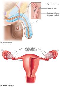
Permanents Methods: Vasectomy, tubal ligation, tubal implants.
Method | Perfect Use Failure Rate (%) | Typical Use Failure Rate (%) |
|---|---|---|
Abstinence | 0 | Unknown |
Withdrawal | 4 | 27 |
Condoms | 2–4 | 15–21 |
Oral contraceptives | 0.3 | 1–2 |
IUD | 0.6–0.8 | 0.2–0.8 |
Vasectomy | 0.1 | 0.15 |
No birth control | 85 | 85 |
Sexually Transmitted Infections (STIs)
Bacterial/Parasitic: Chlamydia, gonorrhea, syphilis, trichomoniasis.
Viral: Human papillomavirus (HPV), genital herpes.
STIs can cause infertility, reproductive disorders, and complications in newborns.
Summary Table: Key Differences Between Spermatogenesis and Oogenesis
Characteristic | Spermatogenesis | Oogenesis |
|---|---|---|
Time of onset | Begins at puberty | Begins before birth |
Number of cells produced | Millions daily | One per month |
Result of meiosis | Four haploid spermatids | One ovum, two polar bodies |
When process ends | Until death | Until menopause |
Additional info: These notes provide a comprehensive overview of the structure, function, and regulation of the male and female reproductive systems, including gametogenesis, hormonal control, and clinical considerations such as infertility, cancer, and contraception. For further details on human development and heredity, see subsequent chapters.
