 Back
BackThe Reproductive System: Structure, Function, and Regulation
Study Guide - Smart Notes
 Tailored notes based on your materials, expanded with key definitions, examples, and context.
The Reproductive System
Overview
The reproductive system ensures the continuation of the species through the production of gametes, fertilization, and support of embryonic development. This chapter focuses on the anatomy and physiology of both male and female reproductive systems, including gametogenesis, hormonal regulation, and the ovarian and testicular cycles.
Male Reproductive System
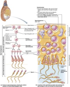
Spermatogenesis
Spermatogenesis is the process by which sperm are produced in the seminiferous tubules of the testes. It involves mitosis, meiosis, and spermiogenesis, resulting in mature spermatozoa.
Mitosis of Spermatogonia: Spermatogonia are stem cells that divide by mitosis. After puberty, each division yields a Type A cell (remains a stem cell) and a Type B cell (becomes a primary spermatocyte).
Meiosis: Primary spermatocytes undergo meiosis I to form two secondary spermatocytes, which then undergo meiosis II to produce four spermatids.
Spermiogenesis: Spermatids elongate, shed excess cytoplasm, and develop a tail, forming mature sperm. Sperm are initially non-motile and gain motility in the epididymis.
Role of Sustentocytes (Sertoli Cells): These support cells form the blood-testis barrier, nourish developing sperm, and regulate the environment for spermatogenesis.
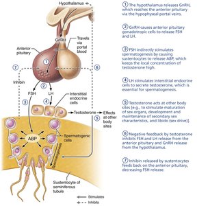
Hormonal Regulation of Male Reproductive Function
The hypothalamic-pituitary-gonadal (HPG) axis regulates spermatogenesis and testosterone production through a series of hormonal interactions.
GnRH: Gonadotropin-releasing hormone from the hypothalamus stimulates the anterior pituitary to release FSH and LH.
FSH: Stimulates sustentocytes to release androgen-binding protein (ABP), maintaining high local testosterone levels for spermatogenesis.
LH: Stimulates interstitial cells to produce testosterone.
Testosterone and Inhibin: Provide negative feedback to the hypothalamus and pituitary to regulate hormone levels.
Testosterone: Mechanism and Effects
Testosterone is a steroid hormone essential for male reproductive function and secondary sex characteristics.
Functions: Stimulates spermatogenesis, growth of reproductive organs, development of secondary sex characteristics (e.g., body hair, deep voice, muscle mass), and libido.
Conversion: In some tissues, testosterone is converted to dihydrotestosterone (DHT) or estradiol for specific effects.
Deficiency: Leads to atrophy of reproductive organs, decreased semen volume, and impaired sexual function.
Female Reproductive System
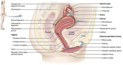
Anatomy of the Female Reproductive System
The female reproductive system is specialized for the production of ova, reception of sperm, fertilization, and support of fetal development. It includes internal and external genitalia.
Ovaries: Produce ova and secrete estrogens and progesterone.
Internal Genitalia: Ovaries, uterine tubes (fallopian tubes), uterus, and vagina.
External Genitalia: Includes structures such as the labia, clitoris, and vestibular glands.
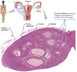
Ovarian Follicles and Oogenesis
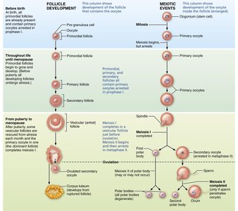
Oogenesis is the process of ovum formation, beginning before birth and continuing until menopause. Ovarian follicles are the functional units of the ovary, each containing an oocyte surrounded by support cells.
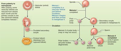
Oogenesis: Oogonia divide by mitosis to form primary oocytes, which are arrested in prophase I until puberty. Each month, some primary oocytes resume meiosis, but only one typically completes ovulation.
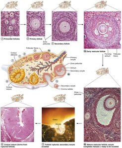
Follicle Development: Follicles mature through primordial, primary, secondary, and vesicular (antral) stages. Ovulation releases a secondary oocyte.
Atresia: Most follicles undergo apoptosis; only a small fraction are ovulated during a woman's reproductive life.
Internal Female Reproductive Organs
The internal organs include the uterine tubes, uterus, and vagina, each with specialized functions in reproduction.
Uterine Tubes: Receive the ovulated oocyte; site of fertilization. Regions include the infundibulum (with fimbriae), ampulla, and isthmus.
Uterus: Muscular organ that receives, retains, and nourishes the fertilized egg. Regions include the body, fundus, isthmus, and cervix.
Vagina: Muscular tube serving as the birth canal, passageway for menstrual flow, and organ of copulation.
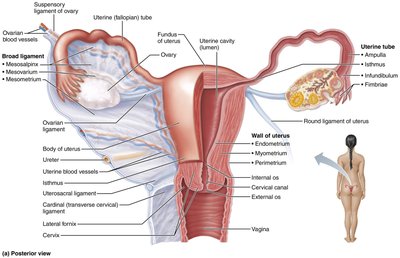
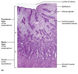
The Uterine Wall and Endometrium
The uterine wall consists of three layers: perimetrium, myometrium, and endometrium. The endometrium is the site of embryo implantation and undergoes cyclic changes during the menstrual cycle.
Functional Layer (Stratum Functionalis): Undergoes cyclic changes and is shed during menstruation.
Basal Layer (Stratum Basalis): Regenerates the functional layer after menstruation.
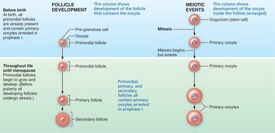
Oogenesis and Follicle Development
Events of Oogenesis
Oogenesis involves the formation of a single mature ovum from each primary oocyte, with the production of polar bodies to discard excess chromosomes.
Primary Oocytes: Arrested in prophase I until puberty.
Secondary Oocytes: Arrested in metaphase II and only complete meiosis if fertilization occurs.
Polar Bodies: Small cells that degenerate, ensuring most cytoplasm remains in the ovum.

Comparison of Oogenesis and Spermatogenesis
Oogenesis and spermatogenesis differ in timing, number of gametes produced, and error rates.
Feature | Male (Spermatogenesis) | Female (Oogenesis) |
|---|---|---|
Total Time to Produce One Gamete | 74 days | 13–50 years |
Occurrence during Lifetime | Puberty to old age | Begins in fetal life, ends in menopause |
Number of Gametes per Meiotic Division | 4 equal-size sperm | 1 large ovum, 2–3 polar bodies |
Number of Gametes per Lifetime | >1 trillion | <500 |
Error Rate (wrong number of chromosomes) | 5% | 20% |
Cells Surrounding Developing Gametes | One sustentocyte sustains many spermatocytes | Many granulosa cells sustain one oocyte |
Stages of Follicle Development
Follicle maturation occurs in two phases: pre-antral (gonadotropin-independent) and antral (gonadotropin-dependent). Follicles progress from primordial to primary, secondary, and vesicular stages, culminating in ovulation and corpus luteum formation.

The Ovarian Cycle
Phases of the Ovarian Cycle
The ovarian cycle is a monthly series of events associated with the maturation of an egg and preparation of the reproductive tract for possible pregnancy.
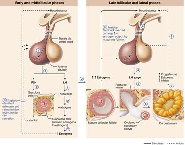
Follicular Phase (Days 1–14): Growth of follicles and increased estrogen secretion; dominant follicle selected for ovulation.
Ovulation (Midcycle): Release of the secondary oocyte from the dominant follicle.
Luteal Phase (Days 15–28): Corpus luteum forms and secretes progesterone and estrogen; degenerates if no pregnancy occurs.
Hormonal Regulation of the Female Reproductive System
Regulation of the Ovarian Cycle
The hypothalamic-pituitary-ovarian axis regulates the ovarian cycle through the interplay of GnRH, FSH, LH, estrogens, and progesterone.
GnRH: Stimulates FSH and LH release from the anterior pituitary.
FSH and LH: Promote follicle growth, estrogen production, and ovulation.
Estrogens and Progesterone: Regulate the uterine cycle and provide feedback to the hypothalamus and pituitary.
Leptin: Adipose-derived hormone that influences the onset of puberty by increasing GnRH release.
Effects of Estrogens and Progesterone
Estrogens and progesterone are critical for female reproductive health and secondary sex characteristics.
Estrogens: Promote oogenesis, follicle growth, and development of secondary sex characteristics (breast development, fat deposition, pelvic widening).
Progesterone: Prepares the endometrium for implantation and supports early pregnancy.
Summary Table: Spermatogenesis vs. Oogenesis
Characteristic | Spermatogenesis | Oogenesis |
|---|---|---|
Time to Produce One Gamete | 74 days | 13–50 years |
Active Years | Puberty to old age | Fetal life to menopause |
Gametes per Meiotic Division | 4 sperm | 1 ovum + 2–3 polar bodies |
Total Gametes per Lifetime | >1 trillion | <500 |
Error Rate | 5% | 20% |
