 Back
BackThe Reproductive System: Structure, Function, and Regulation
Study Guide - Smart Notes
 Tailored notes based on your materials, expanded with key definitions, examples, and context.
The Male Reproductive System
Overview and Main Structures
The male reproductive system is responsible for the production, maturation, and delivery of spermatozoa, as well as the secretion of male sex hormones. It consists of both internal and external organs, each with specialized functions.
Internal organs: Testes, epididymis, vas deferens, seminal vesicles, ejaculatory ducts, prostate gland, urethra, bulbo-urethral glands
External organs: Penis, scrotum
Testes
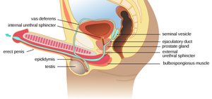
The testes are the primary male reproductive organs, located within the scrotum. They contain seminiferous tubules, which are the site of spermatozoa production (spermatogenesis). The scrotum maintains the testes at a temperature approximately 1°C below core body temperature, which is essential for normal sperm development.
Epididymis
The epididymis is a long, coiled duct (about 6 meters) attached to the posterior surface of each testis. It consists of a head, body, and tail. Sperm mature and are stored here for up to 42 days before being phagocytosed if not ejaculated. The epididymis is continuous with the vas deferens.
Vas Deferens (Ductus Deferens)
The vas deferens is a muscular tube (about 45 cm long) that transports sperm from the epididymis to the ejaculatory duct. It runs anterior to the pubic bone and ends behind the urinary bladder, uniting with the duct from the seminal vesicle just outside the prostate gland.
Seminal Vesicles and Ejaculatory Ducts
The seminal vesicles are tubular glands that secrete about 60% of the seminal fluid, providing nutrients and a transport medium for sperm. The duct from each seminal vesicle joins the vas deferens to form the ejaculatory duct, which passes through the prostate gland and merges with the urethra.
Prostate Gland
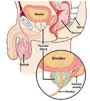
The prostate gland surrounds the first part of the urethra and contributes about 30% of the seminal fluid. Its secretions assist sperm motility and help neutralize vaginal acidity, enhancing sperm survival.
Bulbo-urethral Glands and Urethra
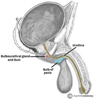
The bulbo-urethral glands (Cowper's glands) secrete a lubricating mucus into the urethra. The male urethra is about 20 cm long and serves as a common passage for urine and semen, ending at the external urethral orifice. It is divided into prostatic, membranous, and penile sections.
Penis
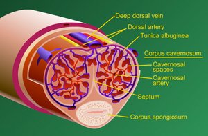
The penis is the external organ of copulation, consisting of a root, shaft, and glans penis (usually covered by the foreskin or prepuce). It contains three columns of erectile tissue: two corpora cavernosa and one corpus spongiosum (surrounding the urethra). These tissues fill with blood during erection.
Sex Cells and Chromosomes
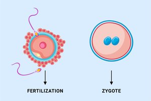
Male and female gametes (sperm and ova) each contain half the normal number of chromosomes. Fertilization restores the diploid number, forming a zygote. Males have XY sex chromosomes, females have XX.
Hormonal Regulation of Male Reproduction
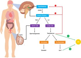
The hypothalamus and anterior pituitary regulate testicular function via two key hormones:
Follicle Stimulating Hormone (FSH): Stimulates spermatogenesis in the seminiferous tubules.
Luteinizing Hormone (LH): Stimulates Leydig cells to produce testosterone.
Testosterone
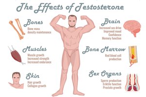
Testosterone is essential for sperm maturation, maintenance of accessory organs, development of secondary sexual characteristics, and stimulation of metabolic processes (e.g., protein synthesis, muscle and bone growth). It also influences brain development, sexual drive, and behaviors.
Spermatozoa Structure
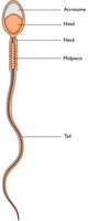
Sperm cells have a head (containing the nucleus and acrosome), a midpiece (rich in mitochondria), and a tail (flagellum) for motility.
The Female Reproductive System
Overview and Main Structures
The female reproductive system is responsible for the production of ova, reception of sperm, fertilization, and support of fetal development. It includes both internal and external organs.
Internal organs: Ovaries, fallopian tubes, uterus, vagina
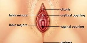
External organs (vulva): Clitoris, labia majora, labia minora, hymen, perineum
The Breasts (Mammary Glands)
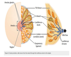
The mammary glands are accessory organs adapted for milk production (lactation). The areola surrounds the nipple, and Montgomery’s tubules secrete lubricating substances to protect the skin.
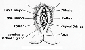
External Genitalia (Vulva)
The vulva includes the clitoris, labia majora, labia minora, hymen, and perineum. These structures protect the internal genital organs and play a role in sexual arousal.
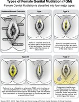
Female Genital Mutilation (FGM)
FGM refers to procedures that intentionally alter or cause injury to the female genital organs for non-medical reasons. It is a violation of human rights and has significant health consequences.
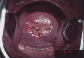
Internal Organs and Structures
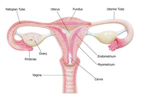
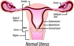
The main internal organs are the uterus, ovaries, fallopian tubes, and vagina. The uterus is a hollow, muscular organ that supports fetal development. The cervix is the lower part of the uterus, separating it from the vagina.
Vagina
The vagina is a muscular tube lined with mucous membrane, extending from the cervix to the external genitalia. It serves as the organ of copulation and the birth canal.
Fallopian Tubes
Each fallopian tube ends in finger-like fimbriae that help waft the egg from the ovary into the tube. Fertilization typically occurs in the distal third of the tube.
edopic pregancy can occur here
Uterus/ womb
The uterus consists of the body, fundus, and cervix. It maintains an environment suitable for implantation and development of a fertilized egg. If fertilization does not occur, the endometrial lining is shed during menstruation. Oxytocin stimulates uterine contractions post-pregnancy. endometriosis occurs here
prolaps uterus
Ovaries and Oogenesis
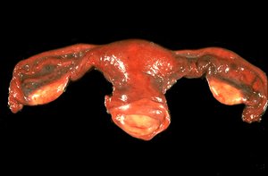
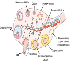
The ovaries are oval organs that store and produce eggs (ova) through oogenesis. Each ovary contains thousands of follicles, each holding an immature oocyte. Ovaries also secrete estrogen and progesterone. one egg amount unless hormone inbalance
Ovarian Follicles and Ovulation
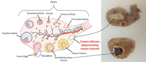
Females are born with all the follicles they will ever have. At puberty, follicle development resumes, and typically one egg matures and is released each month (ovulation). Only 400–500 eggs are ovulated in a woman’s lifetime.
The Ovarian Cycle
The ovarian cycle is divided into three phases:
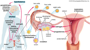
Follicular Phase (Days 1–14): FSH stimulates follicle growth.
Ovulation (Day 14): LH surge triggers release of the mature oocyte./ egg ( with help of estrogen)
Luteal Phase (Days 14–28): The ruptured follicle becomes the corpus luteum, which secretes progesterone to prepare the endometrium for implantation.
The Menstrual Cycle
The menstrual cycle averages 28 days and consists of three phases:
Menstrual Phase (Days 1–5): The endometrial lining is shed, resulting in menstrual flow (average blood loss: 50–100 mL).
Proliferative Phase (Days 6–14): The endometrium regenerates and thickens under the influence of estrogen.
Secretory Phase (Days 15–28): Progesterone from the corpus luteum prepares the endometrium for implantation. If fertilization does not occur, the corpus luteum degenerates, hormone levels fall, and menstruation begins.
Fertilization
The oocyte is viable for 12–24 hours after ovulation, while sperm can fertilize for 24–48 hours after ejaculation. Fertilization restores the diploid chromosome number, forming a zygote.
Key Terms and Concepts
Corpus Luteum: The structure formed from the ruptured follicle after ovulation; secretes progesterone.
Endometrium: The inner lining of the uterus, shed during menstruation.
FSH (Follicle Stimulating Hormone): Stimulates growth of ovarian follicles and spermatogenesis.
LH (Luteinizing Hormone): Triggers ovulation and stimulates testosterone production in males.
Summary Table: Male vs. Female Reproductive Structures
Male Structure | Function | Female Structure | Function |
|---|---|---|---|
Testes | Produce sperm and testosterone | Ovaries | Produce ova, estrogen, progesterone |
Epididymis | Sperm maturation/storage | Fallopian Tubes | Transport ova; site of fertilization |
Vas Deferens | Transports sperm | Uterus | Supports fetal development |
Penis | Copulation, sperm delivery | Vagina | Copulation, birth canal |
