 Back
BackThe Respiratory System: Functional Anatomy and Physiology
Study Guide - Smart Notes
 Tailored notes based on your materials, expanded with key definitions, examples, and context.
The Respiratory System
Overview and Major Functions
The respiratory system is essential for supplying the body with oxygen (O2) for cellular respiration and removing carbon dioxide (CO2), a waste product of metabolism. It also plays roles in olfaction and speech, and is closely linked with the circulatory system for gas transport.
Oxygen Supply: Delivers O2 to tissues for energy production.
CO2 Removal: Eliminates CO2 produced by cells.
Other Functions: Olfaction (smell) and speech.
Four Processes of Respiration
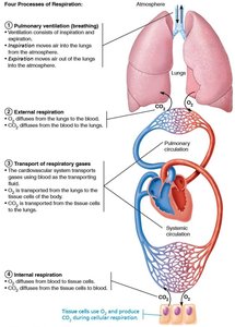
Respiration involves four integrated processes, divided between the respiratory and circulatory systems:
Pulmonary Ventilation: Movement of air into and out of the lungs (breathing).
External Respiration: Exchange of O2 and CO2 between lungs and blood.
Transport of Respiratory Gases: Circulatory system transports O2 and CO2 in blood.
Internal Respiration: Exchange of O2 and CO2 between systemic blood vessels and tissues.
Functional Anatomy of the Respiratory System
Major Organs
The respiratory system is divided into upper and lower regions, each with distinct structures and functions.
Upper Respiratory: Nose, nasal cavity, paranasal sinuses, pharynx
Lower Respiratory: Larynx, trachea, bronchi and branches, lungs, alveoli
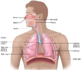
Table: The Upper Respiratory System
Structure | Description, General and Distinctive Features | Function |
|---|---|---|
Nose (external nose and nasal cavity) | External portion supported by bone and cartilage. Internal cavity divided by nasal septum and lined with mucosa. Roof of nasal cavity contains olfactory epithelium. | Produces mucus, filters, warms, and moistens incoming air; resonance chamber for speech; receptors for smell. |
Paranasal Sinuses | Mucosal-lined, air-filled cavities in cranial bones surrounding nasal cavity. | Lighten skull; may also warm, moisten, and filter incoming air. |
Pharynx | Passageway connecting nasal cavity to larynx and oral cavity to esophagus; subdivisions: nasopharynx, oropharynx, laryngopharynx. Houses tonsils (lymphoid tissue masses involved in protection against pathogens). | Passageway for air and food; facilitates exposure of immune system to inhaled antigens. |
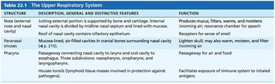
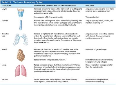
Table: The Lower Respiratory System
Structure | Description, General and Distinctive Features | Function |
|---|---|---|
Larynx | Attaches to pharynx and trachea. Framework of cartilage and dense connective tissue. Opening (glottis) can be closed by epiglottis or vocal folds. | Voice production; provides patent airway; prevents food from entering lower respiratory tract. |
Trachea | Flexible tube running from larynx and dividing inferiorly into two main bronchi. Walls contain C-shaped cartilage rings. | Air passageway; cleans, warms, and moistens incoming air. |
Bronchial Tree | Consists of right and left main bronchi, which subdivide within lungs to form lobar, segmental, and smaller bronchi. Walls contain cartilage and smooth muscle. | Air passageways connecting trachea with alveoli; cleans, warms, and moistens incoming air. |
Alveoli | Microscopic chambers at terminal of bronchial tree. Walls of simple squamous epithelium underlain by basement membrane. | Main sites of gas exchange; surfactant reduces surface tension; helps prevent lung collapse. |
Lungs | Pair of organs flanking mediastinum in thorax. Composed of alveoli and respiratory passageways. | House respiratory passages smaller than main bronchi; alveoli are sites of gas exchange. |
Pleurae | Serous membranes. Parietal pleura lines thoracic cavity; visceral pleura covers external lung surfaces. | Produce lubricating fluid and compartmentalize lungs. |
Upper Respiratory System
The Nose and Paranasal Sinuses
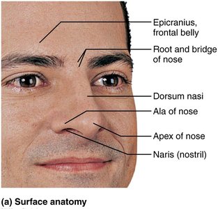
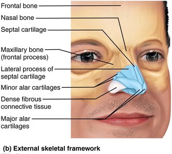
The nose is the only external portion of the respiratory system and serves as the primary entry point for air. It is divided into the external nose and the nasal cavity.
Functions: Provides airway, moistens and warms air, filters and cleans air, serves as a resonating chamber for speech, houses olfactory receptors.
External Nose: Includes root, bridge, dorsum nasi, apex, and nostrils (nares).
Structure: Formed by nasal and frontal bones, maxillary bones, and plates of hyaline cartilage.
Nasal Cavity
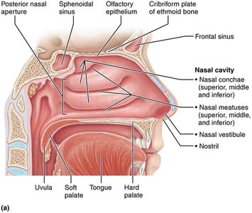
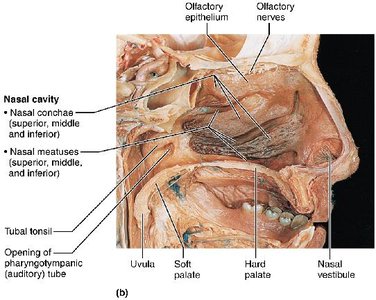
The nasal cavity lies within and posterior to the external nose, divided by the nasal septum. It is lined with mucous membranes and contains structures for filtering, warming, and moistening air.
Septum: Formed by septal cartilage, vomer bone, and perpendicular plate of ethmoid bone.
Roof: Formed by ethmoid and sphenoid bones.
Floor: Formed by hard and soft palates.
Nasal Vestibule: Lined with vibrissae (hairs) for filtering coarse particles.
Mucosa: Olfactory mucosa (superior region) and respiratory mucosa (pseudostratified ciliated columnar epithelium with goblet cells).
Nasal Conchae and Paranasal Sinuses
Nasal conchae are scroll-like, mucosa-covered projections that increase mucosal area and enhance air turbulence. Paranasal sinuses surround the nasal cavity and help lighten the skull, secrete mucus, and warm/moisten air.
Conchae: Superior, middle, and inferior sections; create grooves called meatuses.
Functions: Filter, heat, and moisten air during inhalation; reclaim heat and moisture during exhalation.
Paranasal Sinuses: Located in frontal, sphenoid, ethmoid, and maxillary bones.
Clinical Note: Rhinitis and Sinusitis
Rhinitis is inflammation of the nasal mucosa, which can spread to the throat, chest, tear ducts, and sinuses, causing sinusitis and sinus headaches due to blockage and vacuum formation.
The Pharynx
Structure and Regions
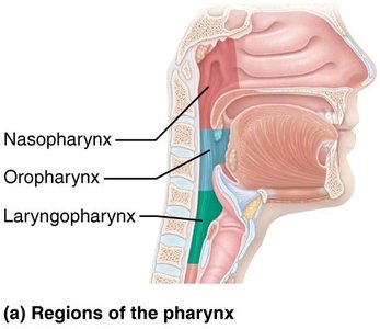
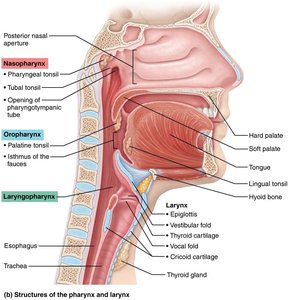
The pharynx is a funnel-shaped muscular tube connecting the nasal cavity and mouth to the larynx and esophagus. It is divided into three regions:
Nasopharynx: Air passageway posterior to nasal cavity; lined with pseudostratified columnar epithelium; contains pharyngeal tonsils and pharyngotympanic tubes.
Oropharynx: Passageway for food and air; lined with stratified squamous epithelium; contains palatine and lingual tonsils.
Laryngopharynx: Passageway for food and air; lined with stratified squamous epithelium; continuous with esophagus.
Clinical Note: Adenoid Enlargement
Swollen adenoids can block air passage in the nasopharynx, forcing mouth breathing and reducing air conditioning. Chronic enlargement may disturb speech and sleep.
Lower Respiratory System
Larynx
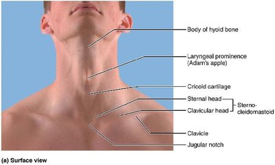
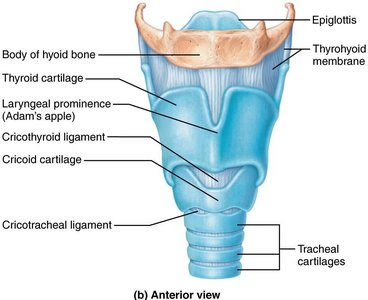
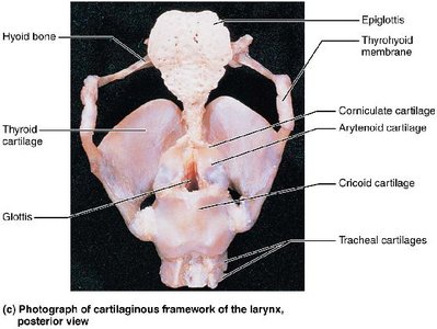
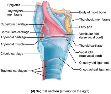
The larynx (voice box) extends from the third to sixth cervical vertebra and attaches to the hyoid bone. It provides a patent airway, routes air and food, and houses vocal folds for voice production.
Framework: Nine cartilages (mostly hyaline), including thyroid, cricoid, arytenoid, cuneiform, corniculate, and epiglottis (elastic cartilage).
Epiglottis: Covers laryngeal inlet during swallowing; covered in taste bud-containing mucosa.
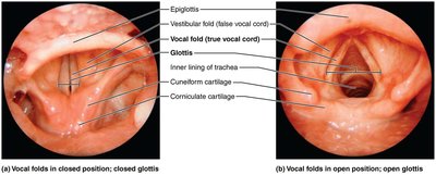
Vocal Folds: True vocal cords; vibrate to produce sound; glottis is the opening between folds.
Vestibular Folds: False vocal cords; help close glottis during swallowing.
Epithelium: Superior portion is stratified squamous; inferior to vocal folds is pseudostratified ciliated columnar.
Voice Production and Sphincter Function
Speech: Intermittent release of expired air during glottis opening/closing.
Pitch: Determined by length and tension of vocal cords.
Loudness: Depends on force of air.
Sphincter Function: Vocal folds can act as a sphincter (e.g., Valsalva's maneuver).
Clinical Note: Laryngitis
Laryngitis is inflammation of the vocal folds, causing hoarseness or loss of voice, often due to infection, overuse, dry air, or irritants.
Trachea
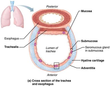
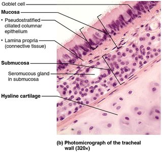
The trachea (windpipe) is a flexible tube extending from the larynx into the mediastinum, dividing into two main bronchi. Its wall has three layers: mucosa, submucosa, and adventitia.
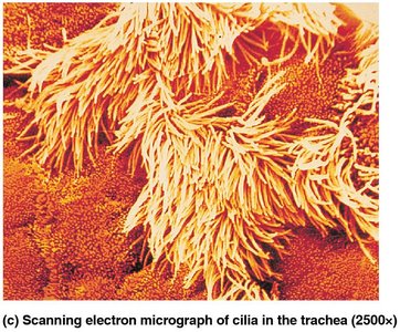
Mucosa: Ciliated pseudostratified epithelium with goblet cells.
Submucosa: Connective tissue with seromucous glands and C-shaped cartilage rings.
Adventitia: Outermost connective tissue layer.
Trachealis: Smooth muscle connecting cartilage rings; contracts during coughing.
Carina: Last tracheal cartilage; highly sensitive, triggers coughing if contacted.
Clinical Notes: Smoking and Tracheal Obstruction
Smoking: Destroys cilia, making coughing the only way to clear mucus; ciliary function recovers after quitting.
Tracheal Obstruction: Life-threatening; Heimlich maneuver can expel obstructions.
Bronchi and Subdivisions
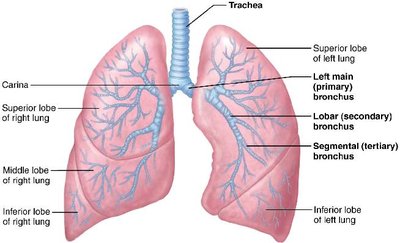
The bronchial tree undergoes 23 orders of branching, transitioning from conducting zone (air transport) to respiratory zone (gas exchange).
Main Bronchi: Right is wider, shorter, more vertical; each enters lung hilum.
Lobar Bronchi: Three on right, two on left; supply lung lobes.
Segmental Bronchi: Branch repeatedly; bronchioles (<1 mm diameter), terminal bronchioles (<0.5 mm diameter).
Conducting Zone Changes: Cartilage rings become plates, elastic fibers replace cartilage, epithelium changes from pseudostratified to cuboidal, smooth muscle increases.
Respiratory Zone Structures
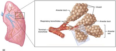
The respiratory zone begins at terminal bronchioles, leading to respiratory bronchioles, alveolar ducts, and alveolar sacs. Alveoli are the main sites of gas exchange.
Alveolar Sacs: Clusters of alveoli (~300 million per lung).
Respiratory Membrane: Thin barrier (~0.5 μm) for gas diffusion; consists of alveolar and capillary walls with fused basement membranes.
Alveolar Cells: Type I (squamous epithelium), Type II (secrete surfactant and antimicrobial proteins).
Other Features: Elastic fibers, pulmonary capillaries, alveolar pores, macrophages.
Lungs and Pleurae
Gross Anatomy of the Lungs
The lungs occupy the thoracic cavity except for the mediastinum. Each lung is divided into lobes and bronchopulmonary segments.
Root: Site of vascular and bronchial attachment.
Hilum: Entry/exit for blood vessels, bronchi, lymphatics, nerves.
Lobes: Right lung (superior, middle, inferior); left lung (superior, inferior, cardiac notch).
Bronchopulmonary Segments: 10 on right, 8–10 on left; each served by its own artery, vein, bronchus.
Lobules: Smallest subdivisions, hexagonal segments served by bronchioles.
Stroma: Elastic connective tissue, makes lungs elastic and spongy.
Blood Supply and Innervation
Pulmonary Circulation: Pulmonary arteries deliver deoxygenated blood; pulmonary veins return oxygenated blood.
Bronchial Circulation: Bronchial arteries supply lung tissue (except alveoli); bronchial veins anastomose with pulmonary veins.
Innervation: Parasympathetic (bronchoconstriction), sympathetic (bronchodilation), visceral sensory fibers.
Pleurae
Pleurae are thin, double-layered serosal membranes dividing the thoracic cavity. Pleural fluid lubricates and assists lung expansion and recoil.
Parietal Pleura: Lines thoracic wall, diaphragm, heart, and between lungs.
Visceral Pleura: Covers external lung surface.
Pleural Fluid: Fills pleural cavity, provides lubrication and surface tension.
Clinical Note: Pleurisy and Pleural Effusion
Pleurisy is inflammation of the pleurae, often due to pneumonia, causing friction and pain. Excess fluid (pleural effusion) may accumulate, hindering breathing.
Additional info: The notes above expand on brief points with academic context, definitions, and examples, and include relevant images and tables to reinforce understanding of respiratory anatomy and physiology.
