 Back
BackThe Respiratory System: Functional Anatomy and Physiology
Study Guide - Smart Notes
 Tailored notes based on your materials, expanded with key definitions, examples, and context.
The Respiratory System: Overview
Introduction
The respiratory system supplies cells with oxygen and eliminates carbon dioxide, playing a critical role in maintaining homeostasis. It is divided into functional anatomy and physiology, each with distinct structures and processes.
Part 1: Functional Anatomy
The Upper Respiratory System
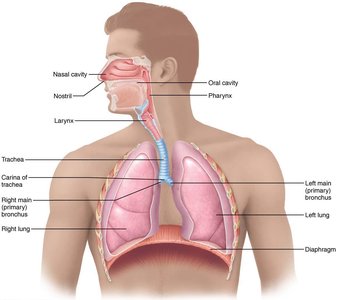
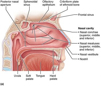
The upper respiratory system includes the nose, nasal cavity, paranasal sinuses, and pharynx. These structures warm, humidify, and filter incoming air, preparing it for entry into the lower respiratory tract.
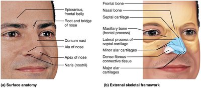
Nose: Provides an airway for respiration, moistens and warms air, filters and cleans incoming air, serves as a resonating chamber for speech, and houses olfactory receptors.
Nasal Cavity: Divided by the nasal septum, lined with mucosa (olfactory and respiratory), and contains nasal conchae to increase surface area for air processing.
Paranasal Sinuses: Located in frontal, maxillary, sphenoid, and ethmoid bones; lighten the skull, warm and moisten air, and produce mucus.
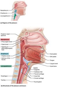
Pharynx: Connects nasal cavity and mouth to the larynx and esophagus; divided into nasopharynx (air passage), oropharynx (air and food passage), and laryngopharynx (air and food passage).
The Larynx
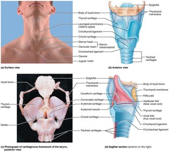
The larynx is a cartilaginous structure that connects the pharynx to the trachea. It provides an open airway, routes food and air into proper passageways, and produces sound via the vocal cords.
Cartilages: Includes thyroid, cricoid, arytenoid, corniculate, cuneiform, and the elastic epiglottis.
Epiglottis: Closes off the larynx during swallowing to prevent food or liquids from entering the airways.
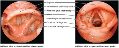
Vocal Folds: Vibrate as air passes over them to produce sound; the glottis is the space between the folds.
The Lower Respiratory System
The lower respiratory system consists of the larynx, trachea, bronchi, and lungs, including the alveoli where gas exchange occurs.
Trachea: Descends from the larynx, supported by C-shaped cartilage rings, lined with ciliated pseudostratified epithelium to propel mucus upward.
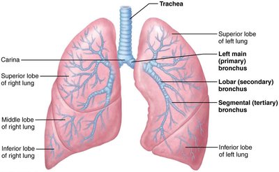
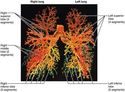
Bronchi: Right and left primary bronchi branch into secondary (lobar) and tertiary (segmental) bronchi, leading to bronchioles.
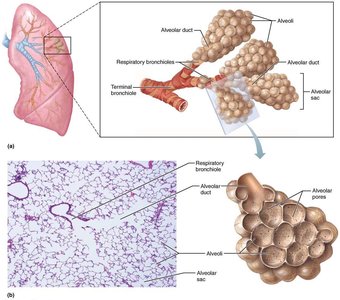
Alveoli: Terminal structures for gas exchange, surrounded by capillaries and elastic fibers.
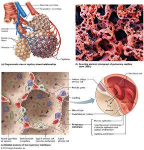
The Lungs and Pleurae
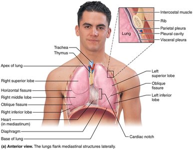
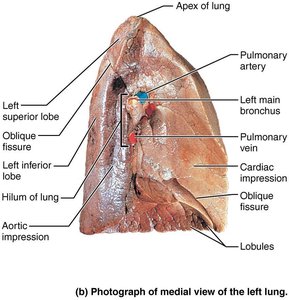
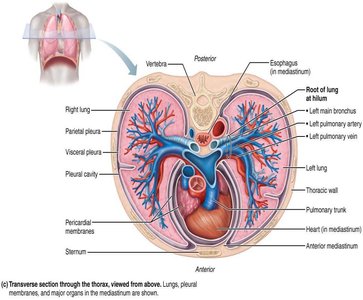
Each lung occupies its own pleural cavity and is divided into lobes and bronchopulmonary segments. The pleurae are double-layered serous membranes that reduce friction and compartmentalize the thoracic cavity.
Lobes: Right lung has three lobes; left lung has two lobes due to the position of the heart.
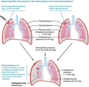
Pleurae: Parietal pleura lines the thoracic wall; visceral pleura covers the lung surface; pleural fluid lubricates the space between layers.
Part 2: Respiratory Physiology
Four Processes of Respiration
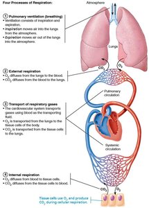
Respiration involves four essential processes: pulmonary ventilation, external respiration, transport of respiratory gases, and internal respiration.
Pulmonary Ventilation: Movement of air into and out of the lungs (breathing).
External Respiration: Exchange of O2 and CO2 between lungs and blood.
Transport of Gases: Movement of O2 and CO2 in the blood via the cardiovascular system.
Internal Respiration: Exchange of O2 and CO2 between blood and tissues.
Pressure Relationships and Pulmonary Ventilation
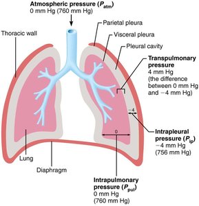
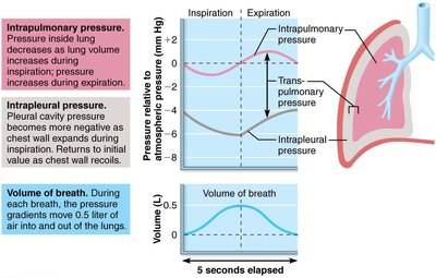
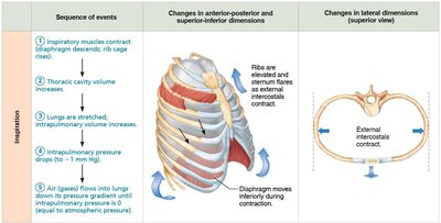
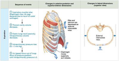
Volume changes in the thoracic cavity cause pressure changes, which drive air movement. Key pressures include atmospheric, intrapulmonary, and intrapleural pressures.
Boyle's Law: At constant temperature, pressure of a gas varies inversely with its volume:
Inspiration: Diaphragm and intercostals contract, increasing thoracic volume and decreasing intrapulmonary pressure, causing air to flow into the lungs.
Expiration: Muscles relax, thoracic volume decreases, intrapulmonary pressure rises, and air flows out.
Airway Resistance, Alveolar Surface Tension, and Lung Compliance
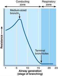
Physical factors influence pulmonary ventilation, including airway resistance, alveolar surface tension, and lung compliance.
Airway Resistance: Friction encountered by air; resistance decreases as airways branch and diameter increases.
Alveolar Surface Tension: Surfactant produced by type II alveolar cells reduces surface tension, preventing alveolar collapse.
Lung Compliance: Measure of lung distensibility; decreased by inflammation, scar tissue, or reduced surfactant.
Respiratory Volumes and Capacities
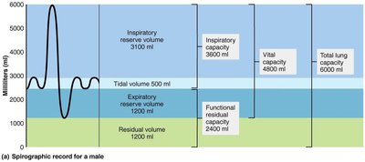
Respiratory volumes and capacities are measured to assess ventilation. Key volumes include tidal volume, inspiratory reserve, expiratory reserve, and residual volume.
Tidal Volume (TV): Amount of air moved in and out during quiet breathing (~500 ml).
Inspiratory Reserve Volume (IRV): Air forcibly inspired beyond TV (2100–3200 ml).
Expiratory Reserve Volume (ERV): Air forcibly expired beyond TV (1000–1200 ml).
Residual Volume (RV): Air remaining after maximal expiration (~1200 ml).
Vital Capacity (VC):
Total Lung Capacity (TLC):
Gas Exchange: External and Internal Respiration
Gas exchange occurs by diffusion, governed by partial pressure gradients and solubility. Dalton's and Henry's laws describe gas behavior in mixtures and liquids.
Dalton's Law: Total pressure of a gas mixture equals the sum of partial pressures of individual gases.
Henry's Law: Gas dissolves in liquid in proportion to its partial pressure.
External Respiration: O2 uptake and CO2 unloading in the lungs; influenced by partial pressure gradients, membrane thickness, and ventilation-perfusion coupling.
Internal Respiration: O2 unloading and CO2 uptake in tissues.
Oxygen and Carbon Dioxide Transport
Oxygen is transported primarily by hemoglobin, while carbon dioxide is carried in three forms: dissolved in plasma, bound to hemoglobin, and as bicarbonate ions.
Oxygen Transport: 98.5% bound to hemoglobin, 1.5% dissolved in plasma.
Carbon Dioxide Transport: 7–10% dissolved in plasma, 20% bound to hemoglobin, 70% as bicarbonate.
Haldane Effect: Deoxygenated hemoglobin carries more CO2; oxygenated hemoglobin releases CO2.
Carbonic Acid-Bicarbonate Buffer:
Control of Respiration
Respiratory centers in the brain stem regulate breathing, with input from chemoreceptors and higher brain centers.
Medullary Centers: Ventral and dorsal respiratory groups control rhythm and depth.
Pontine Centers: Modify breathing rhythm and prevent over-inflation.
Chemoreceptors: Monitor levels of CO2, O2, and H+ in arterial blood; central chemoreceptors in the medulla, peripheral chemoreceptors in the aortic arch and carotid arteries.
Higher Brain Centers: Hypothalamus and cerebral cortex can alter breathing rate and depth.
Respiratory Adjustments: Exercise and High Altitude
Exercise and high altitude require adjustments in respiratory function to meet increased oxygen demands and adapt to lower atmospheric oxygen.
Exercise: Hyperpnea increases depth and rate of respiration; neural factors contribute to changes.
High Altitude: Acute mountain sickness may occur; acclimatization involves increased ventilation, lower hemoglobin saturation, and increased erythropoietin production.
