 Back
The Respiratory System: Functional Anatomy and Physiology
Study Guide - Smart Notes
 Tailored notes based on your materials, expanded with key definitions, examples, and context.
The Respiratory System: Overview
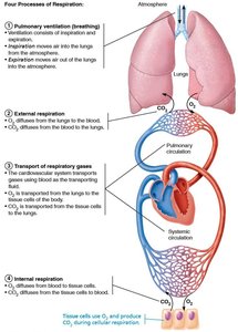
The respiratory system supplies the body with oxygen and removes carbon dioxide, a waste product of cellular metabolism. It is closely linked with the circulatory system and also plays roles in olfaction and speech.
Major Functions: Supplying O2, removing CO2, olfaction, and speech.
Processes of Respiration: Pulmonary ventilation, external respiration, transport of respiratory gases, and internal respiration.
Part 1: Functional Anatomy
Major Organs of the Respiratory System
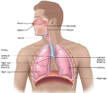
Upper respiratory system: Nose, nasal cavity, paranasal sinuses, pharynx
Lower respiratory system: Larynx, trachea, bronchi and branches, lungs, alveoli
The Upper Respiratory System
The upper respiratory system conditions air by filtering, warming, and humidifying it before it reaches the lower respiratory tract.
Nose and Nasal Cavity: Provides an airway, moistens and warms air, filters particles, serves as a resonance chamber for speech, and houses olfactory receptors.
Paranasal Sinuses: Lighten the skull, produce mucus, and help warm and moisten air.
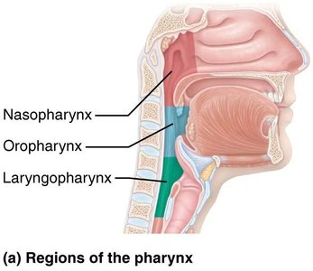
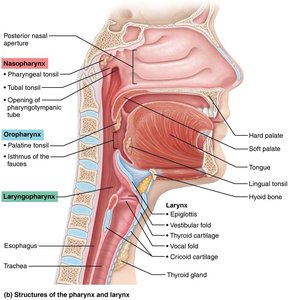
Pharynx: Connects the nasal cavity and mouth to the larynx and esophagus; divided into nasopharynx, oropharynx, and laryngopharynx.
Structure | Description, General & Distinctive Features | Function |
|---|---|---|
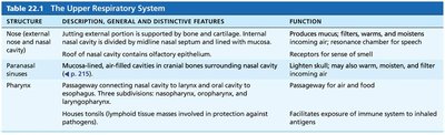
Nose (external nose and nasal cavity) | External portion supported by bone and cartilage. Internal cavity divided by septum and lined with mucosa. Roof contains olfactory epithelium. | Produces mucus, filters, warms, and moistens incoming air; resonance chamber for speech; receptors for smell. |
Paranasal sinuses | Mucosa-lined, air-filled cavities in cranial bones surrounding nasal cavity. | Lighten skull; may also warm, moisten, and filter incoming air. |
Pharynx | Passageway connecting nasal cavity to larynx and oral cavity to esophagus; three subdivisions: nasopharynx, oropharynx, laryngopharynx. Houses tonsils. | Passageway for air and food; facilitates exposure of immune system to inhaled antigens. |
Surface Anatomy and Structure of the Nose
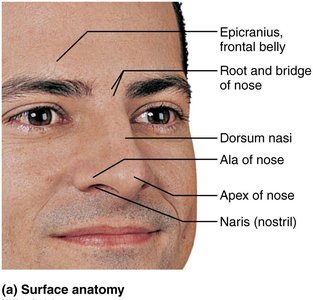
Surface features: Root, bridge, dorsum nasi, apex, ala, and naris (nostril).
Framework: Formed by frontal, nasal, and maxillary bones, and hyaline cartilage.
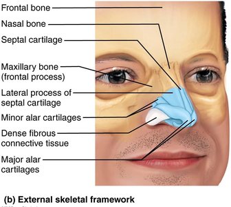
Nasal Cavity
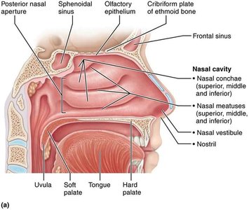
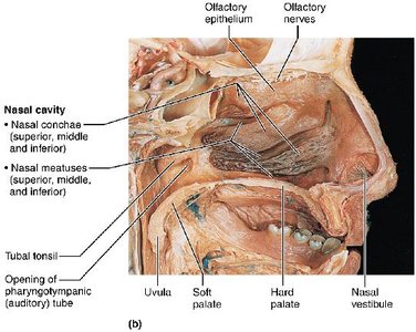
Divided by the nasal septum (cartilage and bone).
Roof formed by ethmoid and sphenoid bones; floor by hard and soft palate.
Lined with vibrissae (hairs) and mucous membranes (olfactory and respiratory mucosa).
Nasal conchae increase surface area and air turbulence, aiding in filtering, warming, and moistening air.
Pharynx
Nasopharynx: Air passageway; contains pharyngeal tonsil and auditory tubes.
Oropharynx: Passageway for food and air; contains palatine and lingual tonsils.
Laryngopharynx: Passageway for food and air; continuous with esophagus.
The Lower Respiratory System
The lower respiratory system conducts air to the lungs and is the site of gas exchange.
Structure | Description, General & Distinctive Features | Function |
|---|---|---|
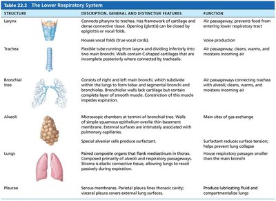
Larynx | Connects pharynx to trachea; framework of cartilage and dense connective tissue; opening (glottis) can be closed by epiglottis or vocal folds. | Air passageway; prevents food from entering lower respiratory tract; voice production. |
Trachea | Flexible tube running from larynx and dividing inferiorly into two main bronchi; walls contain C-shaped cartilage rings. | Air passageway; cleans, warms, and moistens incoming air. |
Bronchial tree | Consists of right and left main bronchi, which subdivide within lungs to form smaller branches; walls contain cartilage and smooth muscle. | Air passageways connecting trachea with alveoli; cleans, warms, and moistens incoming air. |
Alveoli | Microscopic chambers at termini of bronchial tree; walls of simple squamous epithelium overlie thin basement membrane; external surfaces intimately associated with pulmonary capillaries. | Main sites of gas exchange; surfactant reduces surface tension; helps prevent alveoli collapse. |
Lungs | Paired composite organs; right lung has three lobes, left has two; composed primarily of alveoli and respiratory passageways. | House respiratory passages smaller than main bronchi. |
Pleurae | Serous membranes; parietal pleura lines thoracic cavity; visceral pleura covers external lung surface. | Produce lubricating fluid and compartmentalize lungs. |
Larynx Structure and Function
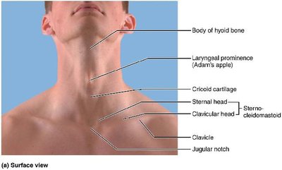
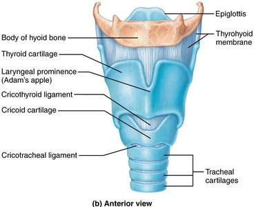
Formed by nine cartilages (thyroid, cricoid, arytenoid, cuneiform, corniculate, epiglottis).
Epiglottis covers the laryngeal inlet during swallowing.
Vocal folds (true vocal cords) produce sound; vestibular folds (false vocal cords) help close the glottis during swallowing.
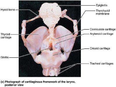
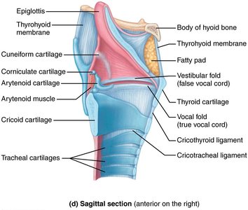
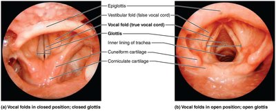
Trachea Structure
Extends from the larynx into the mediastinum, dividing into two main bronchi.
Wall consists of mucosa (ciliated pseudostratified epithelium), submucosa, and adventitia with C-shaped cartilage rings.
Trachealis muscle allows for adjustment of diameter and forceful expulsion of air.
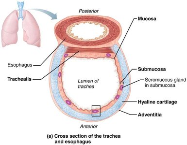
Additional info:
Further sections would cover the bronchial tree, alveoli, lungs, pleurae, and the physiology of breathing, gas exchange, and control of respiration, as outlined in the chapter overview. The images included above are directly relevant to the anatomical and functional explanations provided.
