 Back
BackThe Respiratory System: Gas Transport and Regulation
Study Guide - Smart Notes
 Tailored notes based on your materials, expanded with key definitions, examples, and context.
The Respiratory System: Gas Transport and Regulation
Oxygen Transport in the Blood
Oxygen is transported in the blood primarily by binding to hemoglobin within red blood cells, with a small fraction dissolved directly in plasma. The efficiency of oxygen transport is crucial for meeting the metabolic demands of tissues.
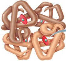
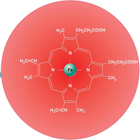
Hemoglobin Structure: Hemoglobin (Hb) consists of four polypeptide chains, each containing an iron-bearing heme group that binds oxygen.
Oxygen Binding: Each hemoglobin molecule can bind up to four O2 molecules, forming oxyhemoglobin (HbO2); when oxygen is released, it becomes reduced hemoglobin (HHb).
Transport Forms:
98.5% of O2 is carried bound to hemoglobin.
~1.5% is dissolved in plasma (O2 is poorly soluble in water).
Oxygen Loading and Unloading: The affinity of hemoglobin for oxygen depends on its saturation level and is influenced by partial pressure of oxygen (PO2), temperature, blood pH, partial pressure of carbon dioxide (PCO2), and the concentration of 2,3-bisphosphoglycerate (BPG).
The Oxygen-Hemoglobin Dissociation Curve
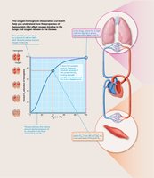
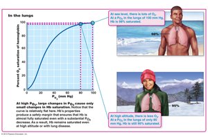
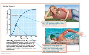
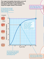
The oxygen-hemoglobin dissociation curve illustrates the relationship between PO2 and hemoglobin saturation. It is sigmoidal, reflecting cooperative binding among hemoglobin's subunits.
Steep Slope: At PO2 values between 10 and 50 mm Hg, small changes in PO2 cause large changes in saturation.
Plateau: At PO2 values between 70 and 100 mm Hg, hemoglobin is nearly fully saturated, even if PO2 drops slightly (important for high altitude or cardiopulmonary disease).
Venous Reserve: Only about 25% of oxygen is unloaded during a single pass through tissues, leaving a reserve for increased demand (e.g., during exercise).
Factors Affecting Hemoglobin Saturation
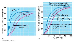
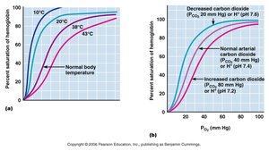
Hemoglobin's affinity for oxygen is modulated by several physiological factors, which shift the dissociation curve to the right (decreased affinity, more O2 unloading) or left (increased affinity, less O2 unloading).
Right Shift (Increased O2 Unloading): Caused by increased temperature, PCO2, H+ (lower pH), and BPG.
Left Shift (Decreased O2 Unloading): Caused by decreased temperature, PCO2, H+, and BPG.
Bohr Effect: Acidosis (increased H+) weakens the Hb-O2 bond, promoting oxygen release where it is most needed (tissues).
Carbon Dioxide Transport in the Blood
Carbon dioxide (CO2) is transported from tissues to the lungs in three main forms:
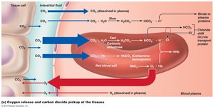
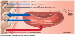
Dissolved in Plasma: 7-10% of CO2 is transported this way.
Bound to Hemoglobin: 20% forms carbaminohemoglobin (CO2 binds to amino acids of Hb, not the heme group).
Bicarbonate Ion (HCO3-): ~70% is converted to bicarbonate in RBCs via the enzyme carbonic anhydrase. The reaction is:
Chloride Shift: Bicarbonate ions diffuse into plasma, and chloride ions move into RBCs to maintain electrical neutrality.
CO2 and Blood pH Regulation
CO2 levels are closely linked to blood pH through the carbonic acid-bicarbonate buffer system. Respiration plays a major role in maintaining acid-base balance.
Slow, Shallow Breathing: CO2 accumulates, carbonic acid increases, and blood pH drops (acidosis).
Rapid, Deep Breathing: CO2 is expelled, carbonic acid decreases, and blood pH rises (alkalosis).
Buffering: H+ ions are buffered by hemoglobin and plasma proteins; HCO3- acts as an alkaline reserve.
Nervous System Control of Respiration
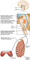
Respiratory rhythm is generated and regulated by centers in the medulla oblongata and pons. The two main medullary centers are the ventral respiratory group (VRG) and dorsal respiratory group (DRG).
Ventral Respiratory Group (VRG): Acts as the primary rhythm generator, stimulating inspiratory muscles via the phrenic and intercostal nerves. Expiratory neurons promote passive expiration.
Dorsal Respiratory Group (DRG): Integrates input from peripheral stretch and chemoreceptors; modulates VRG activity.
Normal Breathing Rate: Eupnea (12-15 breaths/min).
Suppression: Overdose of depressants (e.g., morphine, alcohol) can suppress VRG, leading to respiratory arrest.
Chemical Regulation of Respiration
Respiratory rate and depth are primarily regulated by changes in arterial CO2, O2, and pH, detected by central and peripheral chemoreceptors.
CO2 (Most Potent Stimulus): Central chemoreceptors in the medulla respond to changes in brain extracellular fluid pH (reflecting CO2 levels). Increased CO2 (hypercapnia) lowers pH, stimulating increased ventilation (hyperventilation).
O2: Peripheral chemoreceptors in the aortic arch and carotid arteries respond to significant drops in PO2 (<60 mm Hg), augmenting the response to CO2.
pH: Peripheral chemoreceptors also respond to changes in arterial pH (e.g., from lactic acid or ketone bodies), independent of CO2 or O2 levels.
Other Factors Influencing Respiration
Hering-Breuer Reflex: Stretch receptors in the lungs inhibit inspiration to prevent overinflation (protective reflex).
Hypothalamic Controls: Strong emotions, pain, or temperature changes can alter breathing patterns involuntarily.
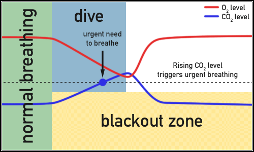
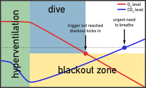
Cortical Controls: Voluntary control over breathing (e.g., holding breath) is possible, but overridden by rising CO2 levels.
Respiratory Adjustments During Exercise
During intense exercise, ventilation increases to meet the elevated demand for oxygen and removal of CO2. This increase (hyperpnea) is matched to metabolic needs and differs from hyperventilation, which is excessive and not driven by metabolic demand.
Phases of Exercise Ventilation:
Rapid increase at onset (anticipation, neural input, proprioceptors, temperature, hormones).
Gradual rise to steady state (linked to CO2 delivery).
Rapid decline post-exercise (removal of stimuli).
Gradual return to baseline (repayment of oxygen debt).
Arterial PO2 and PCO2 remain relatively constant during exercise due to efficient regulation.
Additional info: Inhalation of pure O2 after exercise does not significantly speed recovery because hemoglobin is already nearly fully saturated under normal conditions.
