 Back
BackThe Respiratory System: Structure and Function
Study Guide - Smart Notes
 Tailored notes based on your materials, expanded with key definitions, examples, and context.
The Respiratory System
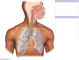
Overview of the Respiratory System and Respiratory Tract
The respiratory system is responsible for the exchange of gases between the atmosphere and the blood, as well as for sound production and regulation of blood pH, volume, and pressure. It is divided into upper and lower respiratory tracts, each with distinct anatomical structures and functions.
Upper respiratory system: Nose, nasal cavity, paranasal sinuses, and pharynx
Lower respiratory system: Larynx, trachea, bronchi, bronchioles, and alveoli
Functions of the Respiratory System
Gas exchange: Provides a surface for oxygen and carbon dioxide exchange between air and blood
Sound production: Involved in verbal communication
Regulation: Assists in controlling blood volume, blood pressure, and body fluid pH
The Respiratory Tract
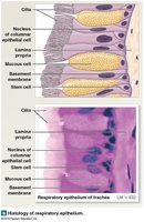
The Respiratory Epithelium
The respiratory tract is lined with specialized epithelium that protects and cleanses the airway.
Pseudostratified ciliated columnar cells: Move mucus and trapped debris upward toward the pharynx (mucociliary escalator)
Stratified squamous cells: Provide protection against abrasion, especially in areas exposed to food and air
Mucus-producing cells: Trap inhaled particles to prevent them from entering the lungs
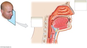
The Upper Respiratory System
The Nose and Nasal Cavity
The nose and nasal cavity filter, warm, and humidify incoming air. They also house olfactory receptors and contribute to resonance in speech.
Dorsum and apex: The bridge and tip of the nose
Nasal bones and cartilages: Form the structure of the nose
Nasal septum: Divides the nasal cavity into right and left portions (formed by the vomer and perpendicular plate of the ethmoid)
Hard and soft palate: Separate the nasal and oral cavities
Nasal conchae: Increase surface area and turbulence for air filtration
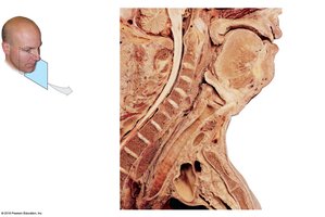
The Pharynx
The pharynx is a muscular tube that serves as a common passageway for air and food, connecting the nasal and oral cavities to the larynx and esophagus.
Nasopharynx: Posterior to the nasal cavity; contains the pharyngeal opening of the auditory tube
Oropharynx: Posterior to the oral cavity; contains the pharyngeal arch and uvula
Laryngopharynx: Inferior region; entrance to the trachea and esophagus
The Lower Respiratory System
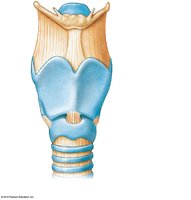
The Larynx
The larynx is a cartilaginous structure that connects the pharynx to the trachea and houses the vocal cords, playing a key role in sound production and airway protection.
Unpaired cartilages: Thyroid (with laryngeal prominence), cricoid, and epiglottis
Paired cartilages: Arytenoid, corniculate, and cuneiform (control opening of the glottis)
Epiglottis: Closes over the glottis during swallowing to prevent food entry into the airway
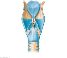
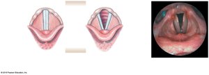
Vocal Folds and Sound Production
Vestibular folds (false vocal cords): Inelastic, do not produce sound
Vocal folds (true vocal cords): Elastic, vibrate to produce sound
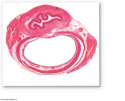
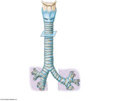
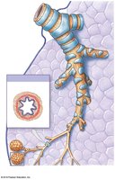
The Trachea
The trachea is a flexible tube that conducts air from the larynx to the bronchi. It is supported by C-shaped rings of hyaline cartilage and bifurcates at the carina into the right and left main bronchi.
Length: Approximately 11 cm; Diameter: 2.5 cm
Cartilage rings: 15–20 C-shaped rings prevent collapse
Trachealis muscle: Allows for constriction and dilation of the trachea

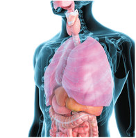
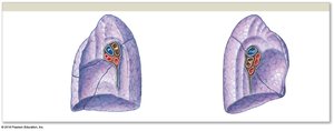
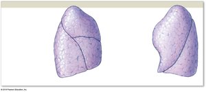
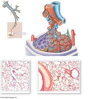
The Main Bronchi and Lungs
The trachea divides into the right and left main bronchi, each entering the lung at the hilum. The lungs are divided into lobes and have distinct surfaces for anatomical orientation.
Right lung: Three lobes (superior, middle, inferior); horizontal and oblique fissures
Left lung: Two lobes (superior, inferior); oblique fissure and cardiac notch
Lung surfaces: Costal, mediastinal, diaphragmatic
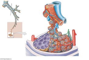
Bronchial Tree and Bronchioles
The bronchial tree consists of branching airways that progressively decrease in size, ending in the alveoli where gas exchange occurs.
Main bronchi: Divide into lobar (secondary) bronchi
Lobar bronchi: Right lung has three, left lung has two
Segmental (tertiary) bronchi: Supply bronchopulmonary segments
Bronchioles: Branch into terminal and respiratory bronchioles
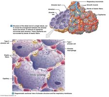
Alveolar Ducts and Alveoli
Alveoli are the primary sites of gas exchange in the lungs. Each alveolus is surrounded by capillaries and elastic fibers, and the alveolar wall forms the blood air barrier.
Type I alveolar cells: Simple squamous cells for gas exchange
Type II alveolar cells: Secrete surfactant to reduce surface tension and prevent collapse
Alveolar macrophages: Phagocytize debris and pathogens
Blood air barrier: Formed by alveolar epithelium, fused basement membrane, and capillary endothelium
Pleural Cavities and Pleural Membranes
Each lung is enclosed by a double-layered serous membrane called the pleura. The pleural cavity contains pleural fluid, which reduces friction during breathing movements.
Visceral pleura: Covers the lung surface
Parietal pleura: Lines the thoracic wall, mediastinum, and diaphragm
Pleural cavity: Space between the layers, filled with pleural fluid
Respiratory Muscles and Pulmonary Ventilation
Pulmonary Ventilation
Pulmonary ventilation is the process of moving air into and out of the lungs, driven by changes in thoracic volume and pressure.
Primary muscles of inhalation: Diaphragm and external intercostals
Normal exhalation: Occurs by relaxation of the diaphragm and external intercostals
Accessory Respiratory Muscles
Muscles aiding inhalation: Serratus anterior, scalenes, pectoralis minor, sternocleidomastoid
Muscles aiding exhalation: Internal intercostals, transversus thoracis, external oblique, internal oblique, rectus abdominis
Summary Table: Major Structures and Functions of the Respiratory System
Structure | Main Function |
|---|---|
Nose/Nasal Cavity | Filters, warms, humidifies air; olfaction |
Pharynx | Passageway for air and food |
Larynx | Sound production; airway protection |
Trachea | Conducts air to bronchi; cleanses air |
Bronchi/Bronchioles | Distribute air to lungs; further cleanse air |
Alveoli | Gas exchange |
Pleura | Reduces friction; compartmentalizes lungs |
