 Back
BackThe Respiratory System: Structure and Function
Study Guide - Smart Notes
 Tailored notes based on your materials, expanded with key definitions, examples, and context.
The Respiratory System
Overview of the Respiratory System
The respiratory system is responsible for the exchange of gases between the body and the environment, primarily oxygen and carbon dioxide. It consists of a series of organs and tissues that facilitate breathing, protect against pathogens, and regulate blood pH.
Key Functions: Gas exchange, filtration of air, warming and moistening of air, vocalization.
Main Structures: Nasal cavity, pharynx, larynx, trachea, bronchi, lungs, alveoli.
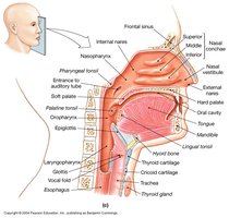

Anatomy of the Respiratory Tract
The respiratory tract is divided into upper and lower regions, each with specialized structures.
Upper Respiratory Tract: Includes the nose, nasal cavity, pharynx, and larynx.
Lower Respiratory Tract: Includes the trachea, bronchi, bronchioles, and lungs.
Nasal Cavity: Filters, warms, and moistens incoming air.
Pharynx: Passageway for air and food; connects nasal cavity to larynx.
Larynx: Contains vocal cords; responsible for sound production.
Trachea: Windpipe; conducts air to the bronchi.
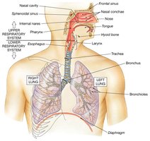
Bronchial Tree
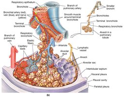
The bronchial tree is a branching system of airways that conducts air from the trachea to the alveoli.
Primary Bronchi: First branches off the trachea, one to each lung.
Secondary Bronchi: Branches to each lobe of the lungs.
Tertiary and Quaternary Bronchi: Further subdivisions, leading to bronchioles.
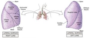
Lobes of the Lungs
The lungs are divided into lobes, each with distinct anatomical features.
Right Lung: Three lobes (superior, middle, inferior).
Left Lung: Two lobes (superior, inferior) and a cardiac notch.
Hilum: Region where bronchi, blood vessels, and nerves enter and exit the lungs.
Respiratory Epithelium
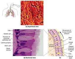
The respiratory epithelium lines the nasal cavity and conducting airways, playing a crucial role in air filtration and protection.
Pseudostratified Ciliated Columnar Epithelium: Contains cilia and goblet cells that trap and move particles out of the airways.
Mucus: Produced by goblet cells; traps dust and pathogens.
Cilia: Move mucus toward the pharynx for removal.
Alveoli and Gas Exchange
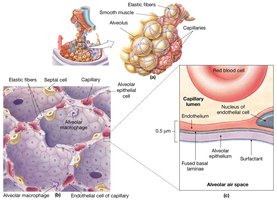
Alveoli are tiny air sacs in the lungs where gas exchange occurs. Their thin, moist lining is essential for efficient oxygen and carbon dioxide transfer.
Structure: Alveoli are surrounded by capillaries and elastic fibers.
Function: Oxygen diffuses from alveoli into blood; carbon dioxide diffuses from blood into alveoli.
Surfactant: Reduces surface tension, preventing alveolar collapse.
Alveolar Macrophages: Remove debris and pathogens.
Air Conditioning in the Nasal Cavity
The nasal cavity conditions incoming air to optimize gas exchange in the alveoli.
Filtration: Hairs and mucus trap particles.
Warming: Blood vessels warm the air.
Moistening: Water vapor humidifies the air.
Muscles of Respiration
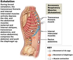
Breathing is facilitated by primary and accessory respiratory muscles.
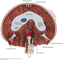
Primary Muscles: Diaphragm and external intercostal muscles.
Accessory Muscles: Sternocleidomastoid, scalene, pectoralis minor, and others assist during forced inhalation and exhalation.
Inhalation: Active process; diaphragm contracts and flattens, increasing thoracic volume.
Exhalation: Usually passive; during forced exhalation, accessory muscles compress the thoracic cavity.

Summary Table: Major Structures and Functions of the Respiratory System
Structure | Function |
|---|---|
Nasal Cavity | Filters, warms, and moistens air |
Pharynx | Passageway for air and food |
Larynx | Sound production, airway protection |
Trachea | Conducts air to bronchi |
Bronchi | Distributes air to lungs |
Lungs | Gas exchange |
Alveoli | Site of gas exchange |
Diaphragm | Main muscle of respiration |
Key Equations
Gas Exchange (Fick's Law): The rate of gas diffusion across the alveolar membrane is given by: Where: A = surface area, D = diffusion coefficient, (P1 - P2) = partial pressure difference, T = thickness of membrane.
Boyle's Law: Describes the relationship between pressure and volume in the lungs:
Clinical Relevance
Diseases: Conditions such as asthma, chronic obstructive pulmonary disease (COPD), and pneumonia affect respiratory function.
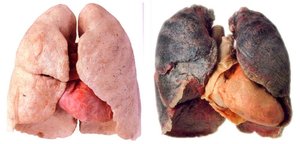
Smoking: Damages respiratory epithelium and alveoli, reducing gas exchange efficiency.
Additional info:
Respiratory system also plays a role in acid-base balance by regulating carbon dioxide levels.
Protective reflexes (coughing, sneezing) help clear airways.
