 Back
BackThe Respiratory System: Structure, Function, and Physiology
Study Guide - Smart Notes
 Tailored notes based on your materials, expanded with key definitions, examples, and context.
The Respiratory System
Overview and Functions
The respiratory system is essential for gas exchange, regulation of blood pH, olfaction (smell), filtering inspired air, sound production, and the removal of small amounts of water and heat. It is structurally and functionally divided into several components that work together to ensure efficient respiration.
Gas Exchange: Oxygen is absorbed and carbon dioxide is expelled.
Blood pH Regulation: By controlling CO2 levels, the respiratory system helps maintain acid-base balance.
Olfaction: Sensory receptors in the nasal cavity detect odors.
Filtration: Air is filtered to remove particles and pathogens.
Sound Production: The larynx produces sounds for speech.
Water and Heat Removal: Small amounts are lost during exhalation.
Structural Organization of the Respiratory System
Major Components
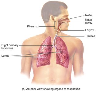
The respiratory system is divided into the upper and lower respiratory tracts, each with distinct anatomical structures and functions.
Upper Respiratory Tract: Includes the nose, nasal cavity, and pharynx.
Lower Respiratory Tract: Includes the larynx, trachea, bronchi, bronchioles, and lungs.
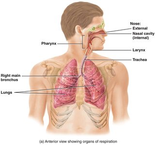
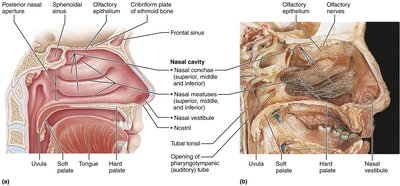
Nose and Nasal Cavity
The nose and nasal cavity are the primary entry points for air. They warm, moisten, and filter incoming air and house olfactory receptors for smell.
Nasal Conchae: Increase surface area and enhance air turbulence for filtration.
Olfactory Epithelium: Contains sensory receptors for smell.
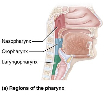
Pharynx
The pharynx is a muscular tube that serves as a passageway for both air and food. It is divided into three regions:
Nasopharynx: Superior region, behind the nasal cavity.
Oropharynx: Middle region, behind the oral cavity.
Laryngopharynx: Inferior region, leading to the larynx and esophagus.
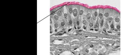
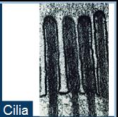
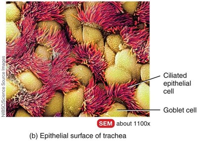
Mucous Membrane and Cilia
The respiratory passageways are lined with a mucous membrane composed of ciliated pseudostratified columnar epithelium. The cilia move mucus and trapped particles toward the pharynx for removal.
Ciliated Cells: Propel mucus and debris out of the respiratory tract.
Goblet Cells: Produce mucus to trap particles.
Lower Respiratory Tract
Larynx
The larynx, or voice box, is involved in sound production and protects the lower airways during swallowing. It contains the vocal cords and several cartilages, including the thyroid and cricoid cartilages.
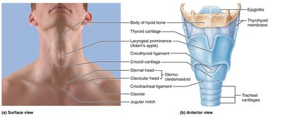
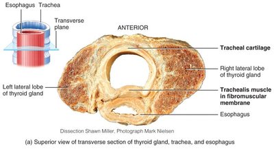
Trachea
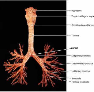
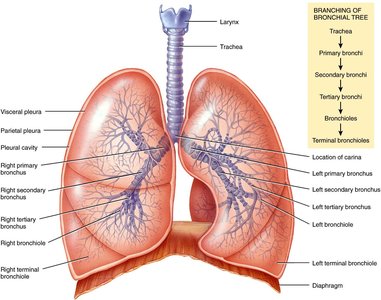
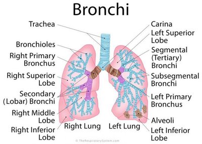
The trachea, or windpipe, is a tube supported by C-shaped cartilaginous rings that maintain airway patency. It divides into the right and left main bronchi at the carina.
Bronchi and Bronchial Tree
The trachea branches into the right and left primary bronchi, which further divide into secondary (lobar), tertiary (segmental), and smaller bronchioles. The bronchial tree ensures air is distributed throughout the lungs.
Bronchioles: Undergo structural changes as they branch; cartilage decreases and smooth muscle increases.
Autonomic Control: Sympathetic stimulation causes bronchodilation; parasympathetic stimulation causes bronchoconstriction.
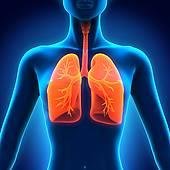
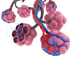
Pulmonary Lobule and Alveoli
The pulmonary lobule is the functional unit of the lung, consisting of a terminal bronchiole, alveolar ducts, alveolar sacs, and alveoli. Alveoli are the primary sites of gas exchange.
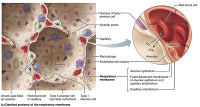
Alveoli: Tiny air sacs surrounded by capillaries where oxygen and carbon dioxide are exchanged.
Respiratory Membrane: Thin barrier (0.5 μm) facilitating rapid gas diffusion.
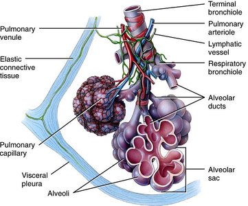
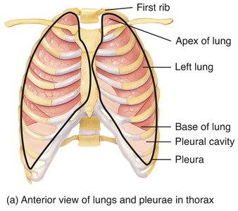
Pleural Membranes
The lungs are enclosed by pleural membranes: the visceral pleura (covers lungs) and parietal pleura (lines thoracic cavity). The pleural cavity contains fluid that reduces friction and allows smooth lung movement during breathing.
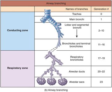
Functional Classification: Conducting and Respiratory Zones
The respiratory system is divided into two functional zones:
Conducting Zone: Includes the nose, pharynx, larynx, trachea, bronchi, and terminal bronchioles. It conducts air to the respiratory zone but does not participate in gas exchange.
Respiratory Zone: Includes respiratory bronchioles, alveolar ducts, and alveoli. This is the site of gas exchange.
Mechanics of Ventilation
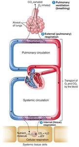
Pulmonary Ventilation
Pulmonary ventilation is the process of moving air into and out of the lungs. It consists of two phases: inhalation (inspiration) and exhalation (expiration).
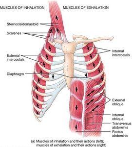
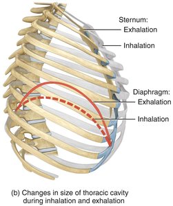
Inhalation: Active process involving contraction of the diaphragm and external intercostal muscles, increasing thoracic volume and decreasing pressure.
Exhalation: Passive during rest, due to elastic recoil of lung tissues and surface tension in alveoli. Becomes active during forceful breathing.
Pressure Changes and Boyle's Law
Boyle's Law states that the pressure of a gas in a closed container is inversely proportional to the volume of the container:
During inhalation, thoracic volume increases and alveolar pressure decreases, causing air to flow into the lungs.
During exhalation, thoracic volume decreases and alveolar pressure increases, causing air to flow out.
Factors Affecting Pulmonary Ventilation
Surface Tension: Alveolar fluid creates surface tension, which must be overcome for alveoli to expand. Surfactant reduces this tension.
Lung Compliance: The ease with which lungs and chest wall expand. High compliance means easy expansion; low compliance means stiff lungs.
Airway Resistance: Resistance to airflow in the airways, primarily in bronchioles. Increased resistance (e.g., in asthma or COPD) makes breathing more difficult.
Gas Exchange: External and Internal Respiration
Gas Laws
Dalton's Law: Each gas in a mixture exerts its own partial pressure. The total pressure is the sum of all partial pressures.
Henry's Law: The amount of gas that dissolves in a liquid is proportional to its partial pressure and solubility.
Example calculation for partial pressure of oxygen in atmospheric air:
External Respiration (Lungs)
Oxygen diffuses from alveolar air (PAlvO2 = 105 mmHg) into pulmonary capillaries (PvO2 = 40 mmHg). Carbon dioxide diffuses from blood (PvCO2 = 45 mmHg) into alveolar air (PAlvCO2 = 40 mmHg).
Internal Respiration (Tissues)
Oxygen diffuses from systemic capillaries (PaO2 = 100 mmHg) into tissue cells (PtO2 = 40 mmHg). Carbon dioxide diffuses from tissue cells (PtCO2 = 45 mmHg) into systemic capillaries (PaCO2 = 40 mmHg).
Respiratory Membrane
The respiratory membrane is a thin barrier (0.5 μm) composed of alveolar epithelium, epithelial basement membrane, interstitial space, capillary basement membrane, and capillary endothelium. It facilitates rapid gas exchange.
Factors Affecting Gas Exchange
Partial pressures of gases
Surface area available for exchange
Diffusion distance (thicker membranes or fluid buildup slow exchange)
Molecular weight and solubility of gases (CO2 diffuses faster than O2 due to higher solubility)
Transport of Gases in the Blood
Oxygen Transport
1.5% of O2 is dissolved in plasma (exerts partial pressure).
98.5% is bound to hemoglobin (Hb) in red blood cells.
Each hemoglobin molecule can bind up to four oxygen molecules.
The amount of oxygen bound to hemoglobin depends on the partial pressure of oxygen (PO2), pH, temperature, PCO2, and 2,3-BPG levels.
Oxygen-Hemoglobin Dissociation Curve
Shows the relationship between PO2 and hemoglobin saturation.
Right shift (decreased affinity): increased acidity, PCO2, temperature, or 2,3-BPG.
Left shift (increased affinity): decreased acidity, PCO2, temperature, or 2,3-BPG.
Carbon Dioxide Transport
7% dissolved in plasma
23% as carbamino compounds (bound to proteins, mainly Hb)
70% as bicarbonate ions (HCO3-)
CO2 + H2O H2CO3 H+ + HCO3-
Control of Breathing
Respiratory Centers
Breathing is controlled by centers in the medulla oblongata and pons. The phrenic nerve stimulates the diaphragm.
Pontine Respiratory Group: Modulates rhythm and prevents over-inflation of the lungs.
Chemoreceptor Regulation
Central and peripheral chemoreceptors monitor CO2, O2, and pH levels.
Increased PCO2 (hypercapnia) stimulates inspiration; decreased PCO2 (hypocapnia) inhibits inspiration.
Central chemoreceptors respond only to PCO2 and H+.
Clinical Note: Hyperventilation decreases PCO2 and allows for longer breath holds (used by swimmers).
Summary Table: Conducting vs. Respiratory Zones
Zone | Structures | Main Function |
|---|---|---|
Conducting Zone | Nose, pharynx, larynx, trachea, bronchi, terminal bronchioles | Conducts air to respiratory zone; filters, warms, and moistens air |
Respiratory Zone | Respiratory bronchioles, alveolar ducts, alveoli | Site of gas exchange |
Additional info: This guide covers the essential structure and function of the respiratory system, including anatomical features, physiological mechanisms, and clinical relevance for ANP college students.
