 Back
BackThe Respiratory System: Structure, Function, and Physiology
Study Guide - Smart Notes
 Tailored notes based on your materials, expanded with key definitions, examples, and context.
The Respiratory System
Overview and Major Functions
The respiratory system is essential for supplying the body with oxygen and removing carbon dioxide, a waste product of metabolism. It consists of a series of organs and structures that facilitate the movement of air, gas exchange, and the transport of respiratory gases in the blood.
Pulmonary ventilation: Movement of air into and out of the lungs (breathing).
External respiration: Gas exchange between the lungs and the blood.
Transport: Movement of oxygen and carbon dioxide between the lungs and tissues via the blood.
Internal respiration: Gas exchange between systemic blood vessels and tissues.
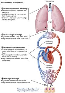
Additional info: Cellular respiration is the process by which cells use oxygen to produce ATP, generating carbon dioxide as a byproduct.
Functional Anatomy of the Respiratory System
Major Respiratory Organs
The respiratory system is divided into upper and lower zones, each with specialized structures and functions.
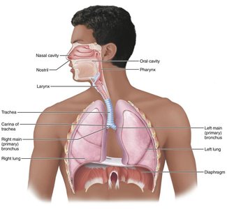
Upper Respiratory Tract
Nose/Nasal cavity: Entry point for air, responsible for filtering, warming, and humidifying incoming air. Contains olfactory receptors for smell.
Paranasal sinuses: Air-filled spaces in cranial bones that lighten the skull and help warm/moisten air.
Pharynx: Muscular tube connecting the nasal cavity and mouth to the larynx and esophagus; divided into nasopharynx, oropharynx, and laryngopharynx.
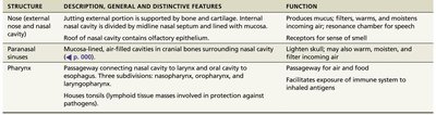
Structure | Description, General and Distinctive Features | Function |
|---|---|---|
Nose (external nose and nasal cavity) | Jutting external portion is supported by bone and cartilage. Internal nasal cavity divided by midline nasal septum and lined with mucosa. Roof of nasal cavity contains olfactory epithelium. | Produces mucus; filters, warms, and moistens incoming air; resonance chamber for speech; receptors for sense of smell. |
Paranasal sinuses | Mucosa-lined, air-filled cavities in cranial bones surrounding nasal cavity. | Lighten skull; may also warm, moisten, and filter incoming air. |
Pharynx | Passageway connecting nasal cavity to larynx and oral cavity to esophagus. Three subdivisions: nasopharynx, oropharynx, and laryngopharynx. Houses tonsils (lymphoid tissue masses involved in protection against pathogens). | Passageway for air and food; facilitates exposure of immune system to inhaled antigens. |
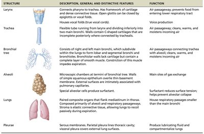
Lower Respiratory Tract
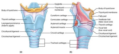
Larynx (voice box): Routes air and food into proper channels; contains vocal cords for sound production.
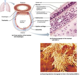
Trachea (windpipe): Flexible tube conducting air to the bronchi; reinforced by C-shaped cartilage rings.
Bronchial tree: Branching system of airways leading from the trachea to the alveoli.
Alveoli: Microscopic air sacs where gas exchange occurs.
Lungs: Paired organs containing the bronchial tree and alveoli; surrounded by pleurae.
Pleurae: Serous membranes that reduce friction and compartmentalize the lungs.
Structure | Description, General and Distinctive Features | Function |
|---|---|---|
Larynx | Connects pharynx to trachea. Has framework of cartilage and dense connective tissue. Open glottis can be closed by epiglottis or vocal folds. | Air passageway; prevents food from entering lower respiratory tract; voice production. |
Trachea | Flexible tube running from larynx and dividing inferiorly into two main bronchi. Walls contain C-shaped cartilage rings. | Air passageway; cleans, warms, and moistens incoming air. |
Bronchial tree | Consists of right and left main bronchus, which subdivide within the lungs to form lobar and segmental bronchi and bronchioles. Walls contain cartilage and smooth muscle. | Air passageways connecting trachea with alveoli; cleans, warms, and moistens incoming air. |
Alveoli | Microscopic chambers at termini of bronchial tree. Walls of simple squamous epithelium overlie thin basement membrane; external surfaces intimately associated with pulmonary capillaries. Produce surfactant. | Main sites of gas exchange; surfactant reduces surface tension; helps prevent alveoli from collapsing. |
Lungs | Paired composite organs that flank mediastinum in thoracic cavity. Composed primarily of alveoli and respiratory passageways. | House respiratory passages smaller than the main bronchi. |
Pleurae | Serous membranes. Parietal pleura lines thoracic cavity; visceral pleura covers external lung surface. | Produce lubricating fluid and compartmentalize lung. |
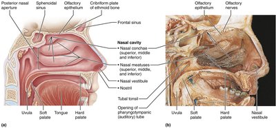
Detailed Anatomy of the Nasal Cavity and Pharynx
The nasal cavity is divided by the nasal septum and contains structures such as the conchae, which increase surface area for warming and humidifying air. The pharynx is divided into three regions: nasopharynx, oropharynx, and laryngopharynx, each with distinct roles in air and food passage.
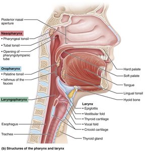
Larynx and Trachea
The larynx is composed of several cartilages, including the thyroid (Adam's apple), cricoid, and epiglottis. The trachea is a flexible tube supported by C-shaped cartilage rings, lined with ciliated epithelium to trap debris.

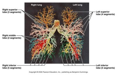
Bronchial Tree and Lungs
The bronchial tree branches extensively within the lungs, ending in alveolar sacs where gas exchange occurs. The right lung has three lobes, while the left has two lobes and a cardiac notch for the heart.
Respiratory Physiology
Mechanics of Breathing
Breathing (pulmonary ventilation) consists of inspiration and expiration, driven by changes in thoracic volume and pressure. Boyle's Law describes the inverse relationship between pressure and volume in the lungs:
Inspiration: Diaphragm and external intercostals contract, increasing thoracic volume and decreasing pressure, causing air to flow in.
Expiration: Muscles relax, thoracic volume decreases, pressure increases, and air flows out.
Additional info: Surfactant produced by Type II alveolar cells reduces surface tension, preventing alveolar collapse.
Gas Laws and Gas Exchange
Dalton's Law: The total pressure of a gas mixture is the sum of the partial pressures of each component gas.
Henry's Law: The amount of gas dissolved in a liquid is proportional to its partial pressure and solubility.
Oxygen and carbon dioxide move across the respiratory membrane by diffusion, driven by partial pressure gradients.
Oxygen and Carbon Dioxide Transport
Oxygen: Transported mainly bound to hemoglobin as oxyhemoglobin; a small amount is dissolved in plasma.
Carbon dioxide: Transported dissolved in plasma, bound to hemoglobin (carbaminohemoglobin), and as bicarbonate ions (HCO3-).
The carbonic anhydrase enzyme catalyzes the conversion of CO2 and H2O to carbonic acid, which dissociates into H+ and HCO3-.
Control of Respiration
Respiratory centers in the medulla and pons regulate the rate and depth of breathing. Chemoreceptors monitor CO2, O2, and pH levels, adjusting ventilation accordingly. The Hering-Breuer reflex prevents over-inflation of the lungs.
Respiratory Volumes and Capacities
Tidal Volume (TV): Volume of air moved in and out with each breath (~500 mL).
Inspiratory Reserve Volume (IRV): Additional air that can be inhaled after a normal inspiration.
Expiratory Reserve Volume (ERV): Additional air that can be exhaled after a normal expiration.
Residual Volume (RV): Air remaining in the lungs after maximal exhalation.
Capacities are combinations of these volumes (e.g., Vital Capacity = TV + IRV + ERV).
Clinical Considerations
Chronic Obstructive Pulmonary Disease (COPD): Includes chronic bronchitis and emphysema; characterized by airflow limitation, dyspnea, and increased risk of infections.
Asthma: Chronic inflammation and bronchospasm leading to wheezing and difficulty breathing.
Tuberculosis: Infectious disease caused by Mycobacterium tuberculosis, forming fibrous nodules in the lungs.
Lung Cancer: Leading cause of cancer death; most cases are related to smoking.
