 Back
BackThe Respiratory System: Structure, Function, and Physiology
Study Guide - Smart Notes
 Tailored notes based on your materials, expanded with key definitions, examples, and context.
The Respiratory System
Overview and Major Functions
The respiratory system is essential for supplying the body with oxygen (O2) and removing carbon dioxide (CO2). It also plays roles in olfaction (sense of smell) and speech production. The system is divided into upper and lower respiratory tracts, each with specialized structures and functions.
Oxygen Supply: Delivers O2 to body tissues for cellular respiration.
Carbon Dioxide Removal: Eliminates CO2, a metabolic waste product.
Olfaction: Houses olfactory receptors for the sense of smell.
Speech: Facilitates sound production via the larynx.
Major Structures of the Respiratory System
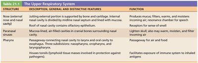
Upper Respiratory Tract
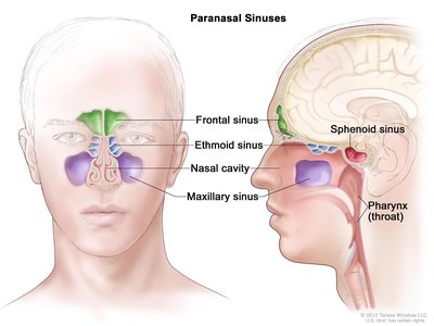
The upper respiratory tract includes the nose, nasal cavity, paranasal sinuses, and pharynx. These structures filter, warm, and moisten incoming air and serve as passageways for airflow.
Structure | Description, Features | Function |
|---|---|---|
Nose/Nasal Cavity | Supported by bone and cartilage; lined with mucosa and vibrissae (hairs) | Filters, warms, moistens air; detects odors; resonance for speech |
Paranasal Sinuses | Air-filled cavities in cranial bones | Lighten skull, secrete mucus, warm/moisten air |
Pharynx | Muscular tube connecting nasal cavity to larynx/esophagus; three regions: nasopharynx, oropharynx, laryngopharynx | Passageway for air and food; houses tonsils for immune defense |
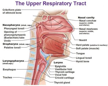
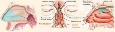
Nasal Cavity and Paranasal Sinuses
Nasal Septum: Divides the nasal cavity; composed of cartilage, vomer, and ethmoid bones.
Olfactory Mucosa: Contains olfactory epithelium for smell.
Respiratory Mucosa: Pseudostratified ciliated columnar epithelium with goblet cells and seromucous glands.
Nasal Conchae: Mucosa-covered projections that increase surface area and enhance air turbulence.
Paranasal Sinuses: Located in frontal, sphenoid, ethmoid, and maxillary bones; lighten skull, secrete mucus, and help warm/moisten air.
Pharynx
The pharynx is a muscular tube with three regions:
Nasopharynx: Posterior to nasal cavity; contains pharyngeal tonsils and pharyngotympanic tubes.
Oropharynx: Posterior to oral cavity; contains palatine and lingual tonsils.
Laryngopharynx: Posterior to epiglottis; leads to larynx and esophagus.
Lower Respiratory Tract
Major Structures and Functions
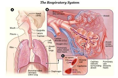
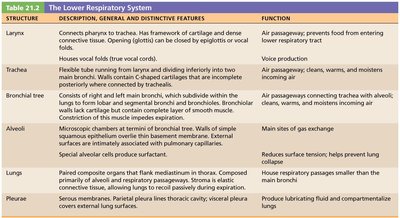
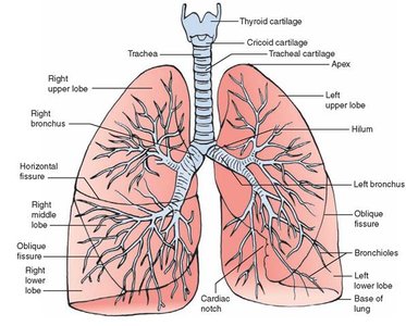
The lower respiratory tract includes the larynx, trachea, bronchi, and lungs. It is divided into the conducting zone (air passageways) and the respiratory zone (site of gas exchange).
Structure | Description, Features | Function |
|---|---|---|
Larynx | Connects pharynx to trachea; contains cartilages and vocal folds | Air passageway, prevents food entry, voice production |
Trachea | Flexible tube with C-shaped cartilage rings | Air passageway; cleans, warms, moistens air |
Bronchial Tree | Branching airways from trachea to alveoli | Air passageways; clean, warm, moisten air |
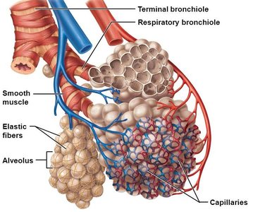
Alveoli | Microscopic air sacs; thin walls | Main site of gas exchange |
Lungs | Paired organs with lobes and stroma | House respiratory passages smaller than main bronchi |
Pleurae | Serous membranes (parietal and visceral) | Produce lubricating fluid, compartmentalize lungs |
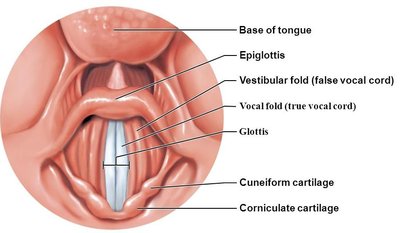
Larynx
Airway: Maintains open passage for air.
Epiglottis: Prevents food from entering the airway during swallowing.
Vocal Folds: Vibrate to produce sound.
Cartilages: Includes thyroid, cricoid, arytenoid, cuneiform, corniculate, and epiglottis.
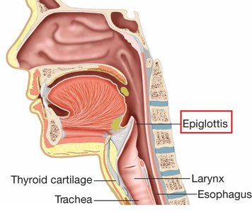
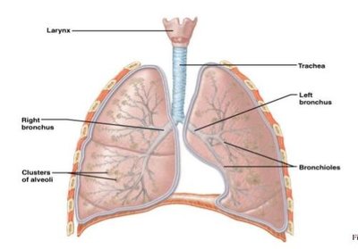
Trachea and Bronchial Tree
Trachea: Windpipe supported by 20 C-shaped cartilage rings; trachealis muscle connects rings posteriorly.
Carina: Sensitive area at the bifurcation of the trachea.
Bronchial Tree: Trachea divides into right and left main bronchi, which branch into smaller bronchi and bronchioles, ending in terminal bronchioles.
Conducting Zone: Passageways for air; cleans, warms, and humidifies.
Respiratory Zone: Terminal bronchioles lead to respiratory bronchioles, alveolar ducts, and alveolar sacs (site of gas exchange).
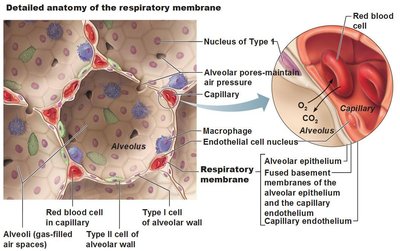
Alveoli and Respiratory Membrane
Alveoli are the primary sites of gas exchange. The respiratory membrane consists of alveolar and capillary walls, allowing efficient diffusion of gases.
Type I Alveolar Cells: Simple squamous cells forming the alveolar wall.
Type II Alveolar Cells: Secrete surfactant to reduce surface tension and antimicrobial proteins.
Respiratory Membrane: Thin barrier (0.5 μm) for rapid gas exchange between alveoli and capillaries.
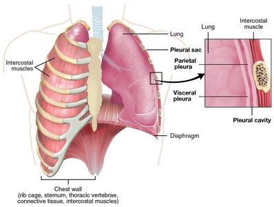
Lungs and Pleurae
Lungs: Right lung has three lobes; left lung has two lobes and a cardiac notch. The hilum is the entry/exit site for bronchi, vessels, and nerves.
Pleurae: Double-layered serous membranes (parietal and visceral) surrounding each lung. Pleural fluid in the pleural cavity reduces friction and allows smooth lung movement.
Mechanics of Breathing (Pulmonary Ventilation)
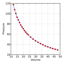
Pressure Relationships and Boyle's Law
Breathing involves two phases: inspiration (inhalation) and expiration (exhalation). Air moves in and out of the lungs due to pressure gradients created by changes in thoracic volume.
Atmospheric Pressure (Patm): Pressure exerted by air outside the body (760 mm Hg at sea level).
Intrapulmonary Pressure (Ppul): Pressure within alveoli; equalizes with atmospheric pressure.
Intrapleural Pressure (Pip): Pressure within pleural cavity; always negative relative to Ppul.
Transpulmonary Pressure: Difference between Ppul and Pip; keeps lungs inflated.
Boyle's Law: The pressure of a gas varies inversely with its volume (if temperature and amount of gas are constant):
Inspiration and Expiration
Inspiration: Active process; diaphragm contracts and flattens, external intercostals lift rib cage, thoracic volume increases, intrapulmonary pressure drops, air flows in.
Expiration: Passive process; inspiratory muscles relax, thoracic volume decreases, intrapulmonary pressure rises, air flows out. Forced expiration involves abdominal and internal intercostal muscles.
Factors Influencing Pulmonary Ventilation
Airway Resistance: Greatest in medium-sized bronchi; increased resistance impedes airflow.
Alveolar Surface Tension: Surfactant (from type II alveolar cells) reduces surface tension, preventing alveolar collapse.
Lung Compliance: The ease with which lungs can expand; decreased by fibrosis, reduced surfactant, or thoracic cage rigidity.
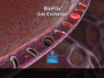
Gas Exchange
External and Internal Respiration
Gas exchange occurs by diffusion across the respiratory membrane (external respiration) and between blood and tissues (internal respiration).
External Respiration: O2 diffuses from alveoli to blood; CO2 diffuses from blood to alveoli. Driven by partial pressure gradients and gas solubility.
Internal Respiration: O2 diffuses from blood to tissues; CO2 diffuses from tissues to blood.
Control of Respiration
Neural and Chemical Regulation
Medullary Respiratory Centers: Ventral respiratory group (VRG) generates rhythm; dorsal respiratory group (DRG) integrates input from stretch and chemoreceptors.
Chemical Factors: Levels of CO2, O2, and pH affect rate and depth of breathing.
Higher Brain Centers: Hypothalamus and cortex can modify respiratory patterns.
Pulmonary Reflexes: Irritant and inflation (Hering-Breuer) reflexes protect the lungs.
Respiratory Pathologies
Chronic Obstructive Pulmonary Disease (COPD)
Emphysema: Destruction of alveolar walls, loss of elasticity.
Chronic Bronchitis: Chronic inflammation and excess mucus production.
Symptoms: Cough, dyspnea, frequent infections; "pink puffers" and "blue bloaters" describe clinical presentations.
Treatment: Bronchodilators, corticosteroids, oxygen therapy, surgery in severe cases.
Asthma
Reversible airway inflammation, bronchospasm, and mucus production.
Triggered by allergens, exercise, or irritants.
Symptoms: Wheezing, coughing, chest tightness, dyspnea.
Tuberculosis (TB)
Infectious disease caused by Mycobacterium tuberculosis.
Symptoms: Fever, night sweats, weight loss, persistent cough, hemoptysis.
Treatment: Long-term antibiotics.
Lung Cancer
Adenocarcinoma: Originates in peripheral lung areas; common in non-smokers.
Squamous Cell Carcinoma: Arises in bronchial epithelium; linked to smoking.
Small Cell Carcinoma: Rapidly metastasizing; originates in primary bronchi.
Treatment: Surgery, radiation, chemotherapy depending on stage and metastasis.
