 Back
The Respiratory System: Structure, Function, and Physiology
Study Guide - Smart Notes
 Tailored notes based on your materials, expanded with key definitions, examples, and context.
The Respiratory System
Overview and Functions
The respiratory system is essential for gas exchange, regulation of blood pH, vocalization, and olfaction. It is divided into upper and lower tracts, each with specialized structures and functions.
Gas Exchange: Intake of O2 and elimination of CO2.
Regulation of Blood pH: By controlling CO2 levels.
Vocalization: Production of sound via the larynx.
Olfactory Receptors: Detection of odors in the nasal cavity.
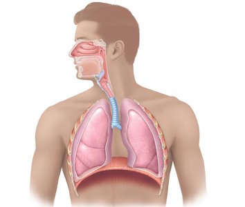
Gross Anatomy of the Respiratory System
Upper Respiratory Tract
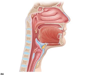
Nose: Provides airway, moistens/warms/filters air, houses olfactory receptors, and adds resonance to voice.
Pharynx: Passageway for air and food, divided into nasopharynx, oropharynx, and laryngopharynx.
Nose Anatomy
External Nose: Entry point for air via external nares (nostrils).
Nasal Cavities: Lined with pseudostratified ciliated columnar epithelium, containing goblet cells and mucus glands for filtration and protection.
Paranasal Sinuses: Located in frontal, maxillary, sphenoid, and ethmoid bones; warm/moisten air, trap debris, and add resonance to voice.
Pharynx Anatomy
Nasopharynx: Air passageway only; lined with pseudostratified ciliated columnar epithelium.
Oropharynx: Common passageway for food and air; lined with stratified squamous epithelium.
Laryngopharynx: Common pathway for food and air; site of divergence of respiratory and digestive systems; stratified squamous epithelium.
Lower Respiratory Tract
Larynx: Maintains open airway, routes food and air, and houses vocal cords for sound production.
Trachea: Conducts air from larynx into thoracic cavity; supported by hyaline cartilage rings.
Bronchi and Lungs: Bronchi branch into bronchioles and alveoli, where gas exchange occurs.
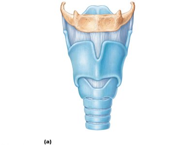
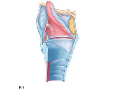
Larynx Anatomy and Function
Thyroid Cartilage: Largest cartilage, contains vocal cords.
Epiglottis: Elastic cartilage flap preventing food from entering airway.
Other Cartilages: Arytenoid and cricoid support vocal folds.
Functions: Separates food and air, produces voice, maintains open airway.
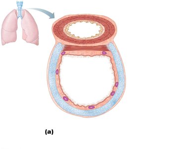
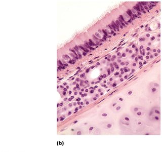
Trachea Histology and Function
Mucosa: Pseudostratified ciliated columnar epithelium with goblet cells for mucus production.
Submucosa: Areolar connective tissue and mucus glands for support and debris trapping.
Hyaline Cartilage: Rings keep airway open.
Adventitia: Areolar connective tissue secures trachea in place.
Function: Air conduction from larynx to thoracic cavity.
Bronchi and Bronchioles
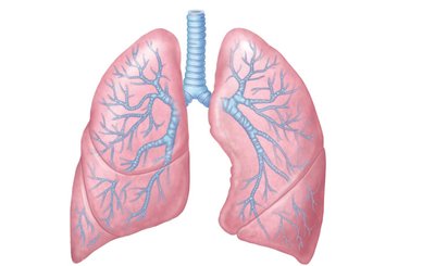
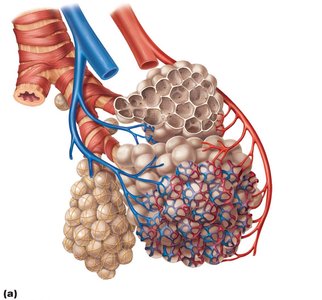
Conducting Zone: Trachea divides into right and left primary bronchi, which branch into smaller bronchi and bronchioles (~25 divisions).
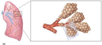
Respiratory Zone: Terminal bronchioles lead to respiratory bronchioles and alveolar sacs (clusters of alveoli).
Bronchioles: Mucosa changes to simple cuboidal epithelium; smooth muscle replaces cartilage, allowing autonomic control (bronchoconstriction/bronchodilation).
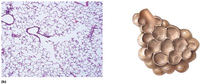
Alveoli and Respiratory Membrane
Function: Site of gas exchange between air and blood.
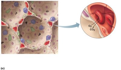
Structure: Single layer of simple squamous epithelium fused to capillary basement membrane (respiratory membrane).
Cell Types:
Type I cells: Form alveolar walls.
Type II cells: Secrete surfactant to reduce surface tension and prevent collapse.
Macrophages: Remove dust and debris.
Changes in the Respiratory Tree
Cartilage rings in trachea are replaced by smooth muscle in bronchioles.
Epithelium transitions from pseudostratified ciliated columnar (nose, sinuses, nasopharynx, trachea, bronchi) to stratified squamous (oropharynx, laryngopharynx) to simple cuboidal (bronchioles) to simple squamous (alveoli).
Smooth muscle in bronchioles is under autonomic control (epinephrine/norepinephrine).
Lungs and Pleura
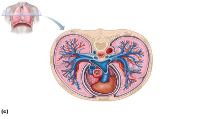
Each lung is in its own cavity lined with pleura (double-layered serous membrane).
Parietal Pleura: Attaches lung to thoracic wall.
Visceral Pleura: Secretes pleural fluid to reduce friction and allow free movement.
Lobes: Left lung has 2 lobes; right lung has 3 lobes.
Processes of Respiration
Types of Respiration
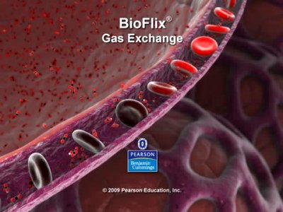
Pulmonary Ventilation: Movement of air in and out of lungs (breathing).
External Respiration: Gas exchange between alveoli and blood.
Internal Respiration: Gas exchange between blood and tissues.
Transport of Gases: Movement of O2 and CO2 in blood (cardiovascular system).
Cellular Respiration: Metabolic reactions producing ATP using O2 and releasing CO2.
External and Internal Respiration
External Respiration: O2 diffuses from alveoli to capillaries; CO2 diffuses from capillaries to alveoli.
Internal Respiration: O2 diffuses from blood to tissues; CO2 diffuses from tissues to blood.
Ventilation Processes
Inhalation: Thoracic cavity volume increases, diaphragm contracts and moves down, external intercostals elevate rib cage, intrapulmonary pressure decreases, air enters lungs.
Exhalation: Thoracic cavity volume decreases, diaphragm and rib muscles relax, intrapulmonary pressure increases, air exits lungs. Passive process dependent on lung elasticity.
Pulmonary Ventilation and Boyle's Law
Ventilation depends on pressure gradients between atmospheric and alveolar pressures.
Boyle's Law: At constant temperature, pressure of a gas varies inversely with its volume.
Factors Affecting Ventilation
Airway Resistance: Increased resistance decreases airflow.
Lung Compliance: Ease of lung expansion.
Lung Elasticity: Ability to recoil (impaired in emphysema).
Alveolar Surface Tension: Surfactant reduces surface tension, preventing collapse.
Diffusion Through the Respiratory Membrane
Thin membrane and large surface area favor rapid gas exchange.
Diffusion is enhanced by increased surface area, thin membrane, high solubility, and steep partial pressure gradients.
Ventilation-perfusion coupling matches airflow and blood flow for optimal exchange.
Clinical Note: Diseases like emphysema, pneumonia, or heart failure can reduce surface area or increase membrane thickness, impairing gas exchange.
Oxygen and Carbon Dioxide Transport
Oxygen Transport and the Bohr Effect
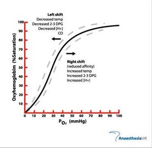
O2 is carried mainly by hemoglobin as oxyhemoglobin.
O2 release from hemoglobin increases with higher CO2, lower pH (Bohr effect), higher temperature, and increased 2,3-BPG.
Bohr Effect: O2 binding affinity to hemoglobin is inversely related to acidity and CO2 concentration.
Carbon Dioxide Transport
CO2 is transported in three forms:
Dissolved in plasma (7%)
Bound to hemoglobin as carbaminohemoglobin (23%)
As bicarbonate ion (HCO3-) (70%)
In red blood cells, carbonic anhydrase catalyzes the reaction:

Chloride shift: HCO3- exits RBCs, Cl- enters to maintain charge balance.
Control of Respiration
Neural and Chemical Regulation
Medulla: Generates and modifies respiratory rate.
Pons: Smooths respiratory rhythm.
Higher Centers: Hypothalamus (sympathetic control), cortex (voluntary control).
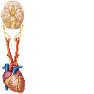
Chemoreceptors: Detect changes in CO2, O2, and pH.
Central chemoreceptors: Brainstem; Peripheral chemoreceptors: Aortic arch and carotid arteries.
CO2 is the most potent regulator; increased CO2 triggers hyperventilation, decreased CO2 triggers hypoventilation.
Respiratory Volumes and Capacities
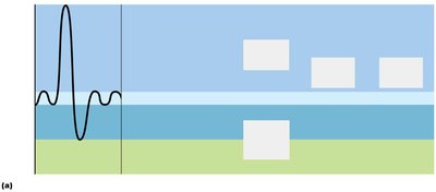
Spirometry and Lung Volumes
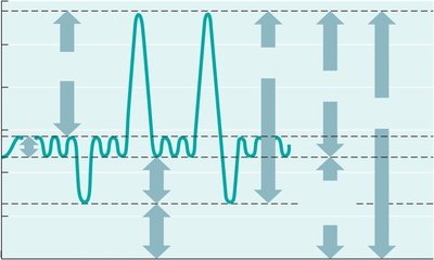
Tidal Volume (TV): Air moved in/out during normal breathing (~500 mL).
Inspiratory Reserve Volume (IRV): Maximal air inhaled above TV (male: 3100 mL, female: 1900 mL).
Expiratory Reserve Volume (ERV): Maximal air exhaled beyond TV (male: 1200 mL, female: 700 mL).
Residual Volume (RV): Air remaining after forced expiration (male: 1200 mL, female: 1100 mL).
Total Lung Capacity (TLC): Maximum air in lungs after maximal inspiration (male: 6000 mL, female: 4200 mL).
Vital Capacity (VC): Maximal air expired after maximal inspiration (male: 4800 mL, female: 3100 mL).
Inspiratory Capacity (IC): Maximal air inspired after normal expiration (male: 3600 mL, female: 2400 mL).
Functional Residual Capacity (FRC): Air remaining after normal expiration (male: 2400 mL, female: 1800 mL).
Measurement | Adult Male | Adult Female | Description |
|---|---|---|---|
Tidal Volume (TV) | 500 mL | 500 mL | Air inhaled/exhaled with each breath at rest |
Inspiratory Reserve Volume (IRV) | 3100 mL | 1900 mL | Max air inhaled after normal inspiration |
Expiratory Reserve Volume (ERV) | 1200 mL | 700 mL | Max air exhaled after normal expiration |
Residual Volume (RV) | 1200 mL | 1100 mL | Air remaining after forced expiration |
Total Lung Capacity (TLC) | 6000 mL | 4200 mL | Max air in lungs after max inspiration |
Vital Capacity (VC) | 4800 mL | 3100 mL | Max air expired after max inspiration |
Inspiratory Capacity (IC) | 3600 mL | 2400 mL | Max air inspired after normal expiration |
Functional Residual Capacity (FRC) | 2400 mL | 1800 mL | Air remaining after normal expiration |
Example: Spirometry is used clinically to assess lung function and diagnose respiratory diseases.
