 Back
The Respiratory System: Structure, Function, and Clinical Correlates
Study Guide - Smart Notes
 Tailored notes based on your materials, expanded with key definitions, examples, and context.
The Respiratory System
Overview and Basic Functions
The respiratory system is essential for supplying the body with oxygen and removing carbon dioxide, a waste product of metabolism. It is composed of a series of organs and tissues that facilitate the movement and exchange of gases between the atmosphere and the bloodstream.
Primary Functions: Supplies oxygen to body tissues and removes carbon dioxide.
Processes of Respiration:
Pulmonary ventilation: Movement of air into and out of the lungs.
External respiration: Gas exchange between air in alveoli and blood in pulmonary capillaries.
Transport of respiratory gases: Movement of gases in the blood between lungs and tissues.
Internal respiration: Gas exchange between systemic blood and tissue cells.
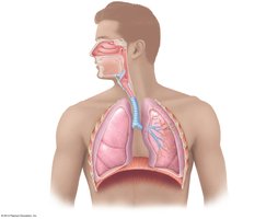
Functional Anatomy of the Respiratory System
Major Organs and Divisions
The respiratory system is divided into the conducting zone and the respiratory zone, each with distinct roles in air passage and gas exchange.
Conducting Zone: Includes the nose, nasal cavity, pharynx, larynx, trachea, bronchi, and terminal bronchioles. These structures filter, warm, and humidify incoming air but do not participate in gas exchange.
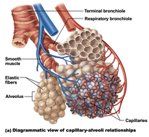
Respiratory Zone: Composed of respiratory bronchioles, alveolar ducts, and alveoli. This is the site of gas exchange.
Organs of the Respiratory System
Nose and Nasal Cavity: Airway for respiration, moistens and warms air, filters particles, and houses olfactory receptors.
Pharynx: Funnel-shaped passageway divided into nasopharynx, oropharynx, and laryngopharynx; serves as a pathway for air and food.
Larynx: Voice production, open airway, routes air and food into proper channels.
Trachea: Windpipe supported by C-shaped cartilage rings.
Bronchi and Bronchioles: Extensive branching passageways leading to alveoli.
Lungs and Alveoli: Main organs of respiration; alveoli are the primary site of gas exchange.
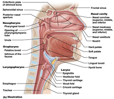
The Nose and Nasal Cavity
Structure and Function
The nose provides an airway for respiration, moistens and warms incoming air, filters inhaled particles, and serves as a resonating chamber for speech. The nasal cavity is divided by the nasal septum and lined with mucous membranes.
Olfactory mucosa: Contains smell receptors near the roof of the nasal cavity.
Respiratory mucosa: Pseudostratified ciliated columnar epithelium with goblet cells; traps and moves particles toward the pharynx.
The Pharynx
Regions and Functions
The pharynx connects the nasal cavity and mouth to the larynx and esophagus. It is divided into three regions:
Nasopharynx: Air passageway; contains pharyngeal tonsil and opening to the auditory tube.
Oropharynx: Passageway for food and air; contains palatine and lingual tonsils.
Laryngopharynx: Passageway for food and air; continuous with the esophagus and larynx.
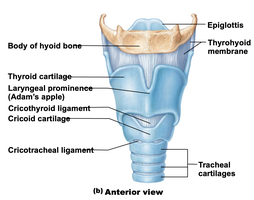
The Larynx
Structure and Function
The larynx, or voice box, is composed of nine cartilages and is responsible for voice production, maintaining an open airway, and routing air and food into the proper channels.
Major Cartilages: Thyroid cartilage (Adam’s apple), cricoid cartilage, arytenoid, corniculate, cuneiform cartilages, and epiglottis.
Vocal Folds: True vocal cords produce sound; vestibular folds (false vocal cords) do not.
Epithelium: Stratified squamous epithelium superiorly, pseudostratified ciliated columnar epithelium inferiorly.
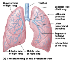
The Trachea and Bronchial Tree
Trachea
The trachea descends from the larynx into the mediastinum and divides into the right and left primary bronchi. It is supported by C-shaped cartilage rings and lined with pseudostratified ciliated columnar epithelium.
Bronchial Tree
The bronchial tree consists of branching airways that decrease in size from the primary bronchi to terminal bronchioles. Tissue composition changes along the tree, with cartilage decreasing and smooth muscle increasing.
Primary (main) bronchi: Enter each lung.
Secondary (lobar) bronchi: Three on the right, two on the left.
Tertiary (segmental) bronchi: Supply bronchopulmonary segments.
Bronchioles: Less than 1 mm in diameter; terminal bronchioles are less than 0.5 mm.
The Respiratory Zone
Alveoli and Gas Exchange
The respiratory zone includes respiratory bronchioles, alveolar ducts, and alveoli. Gas exchange occurs across the respiratory membrane, which consists of alveolar and capillary walls and their fused basal laminae.
Type I alveolar cells: Simple squamous epithelial cells forming the structure of the alveolar wall.
Type II alveolar cells: Secrete surfactant to reduce surface tension and prevent alveolar collapse.
Alveolar macrophages: Remove debris and pathogens from the alveolar surface.
Alveolar pores: Connect adjacent alveoli and equalize air pressure.
Gross Anatomy of the Lungs
Lobes, Fissures, and Pleurae
The lungs are divided into lobes by fissures. The right lung has three lobes (superior, middle, inferior), and the left lung has two lobes (superior, inferior) and a cardiac notch. Each lung is surrounded by a double-layered pleural membrane (parietal and visceral pleura) with a pleural cavity in between.
Hilum: Region where bronchi, blood vessels, lymphatics, and nerves enter and exit the lung.
Pleurae: Reduce friction and compartmentalize the thoracic cavity.
Mechanisms of Ventilation
Inspiration and Expiration
Ventilation consists of two phases: inspiration (inhalation) and expiration (exhalation). These processes depend on changes in thoracic cavity volume and pressure.
Inspiration: Diaphragm contracts and flattens, external intercostal muscles contract, increasing thoracic volume and decreasing pressure, causing air to flow in.
Expiration: Usually passive; diaphragm and intercostals relax, thoracic volume decreases, pressure increases, and air flows out. Forced expiration involves abdominal and internal intercostal muscles.
Key Equation:
(Boyle's Law: Pressure and volume are inversely related in a closed system at constant temperature.)
Neural Control of Ventilation
Respiratory Centers and Chemoreceptors
Breathing is regulated by respiratory centers in the brainstem, primarily the ventral respiratory group (VRG) in the medulla oblongata. Chemoreceptors in the medulla (central) and in the aortic and carotid bodies (peripheral) monitor blood levels of CO2, O2, and pH to adjust ventilation rate.
Clinical Correlates
Disorders of the Lower Respiratory Tract
Bronchial Asthma: Allergic inflammation causing bronchoconstriction and mucus secretion.
Cystic Fibrosis: Inherited disorder leading to thick, sticky mucus that obstructs airways.
Chronic Obstructive Pulmonary Disease (COPD): Includes emphysema (destruction of alveolar walls, loss of elasticity) and chronic bronchitis (excess mucus, productive cough). Most common cause is smoking.
Example: In emphysema, the surface area for gas exchange is greatly reduced, leading to impaired oxygenation of blood.
Disorder | Main Features | Causes |
|---|---|---|
Asthma | Bronchoconstriction, mucus secretion, airway inflammation | Allergens, irritants, stress |
Cystic Fibrosis | Thick, viscous mucus, airway obstruction | Genetic mutation (CFTR gene) |
COPD (Emphysema, Chronic Bronchitis) | Airflow limitation, alveolar destruction, chronic cough | Smoking, air pollution |
Additional info: The respiratory system also plays a role in speech, olfaction, and acid-base balance by regulating CO2 levels in the blood.
