 Back
The Respiratory System: Structure, Function, and Physiology
Study Guide - Smart Notes
 Tailored notes based on your materials, expanded with key definitions, examples, and context.
The Respiratory System
Overview and Functional Anatomy
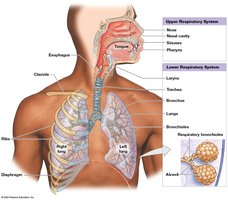
The respiratory system is responsible for the exchange of gases (oxygen and carbon dioxide) between the atmosphere and the blood. It is divided into structural and functional components that facilitate ventilation and gas exchange.
Upper respiratory system: Includes the nose, nasal cavity, paranasal sinuses, and pharynx.
Lower respiratory system: Comprises the larynx, trachea, bronchi, bronchioles, and lungs.
Conducting portion: Series of passageways that conduct air into the lungs (nose to terminal bronchioles).
Respiratory portion: Sites of gas exchange (respiratory bronchioles and alveoli).
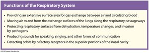
Major Functions of the Respiratory System
Provides an extensive surface area for gas exchange between air and circulating blood.
Moves air to and from the exchange surfaces of the lungs along the respiratory passageways.
Protects respiratory surfaces from dehydration, temperature changes, and invasion by pathogens.
Produces sounds for speaking, singing, and other forms of communication.
Detects odors by olfactory receptors in the nasal cavity.
Histology and Protection of the Respiratory Tract
Respiratory Mucosa and Epithelium
The respiratory tract is lined by a mucosa that varies in structure along its length, providing protection and facilitating its functions.
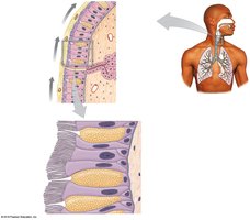
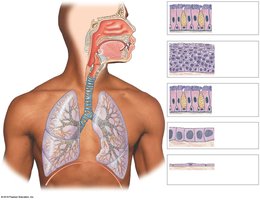
Pseudostratified ciliated columnar epithelium: Lines the nasal cavity, upper pharynx, trachea, bronchi, and large bronchioles. Cilia move debris toward the pharynx (mucociliary escalator).
Stratified squamous epithelium: Lines the lower pharynx, providing protection against abrasion.
Cuboidal epithelium: Found in smaller bronchioles.
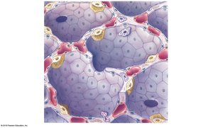
Simple squamous epithelium: Lines the alveoli, facilitating gas exchange.
Cystic Fibrosis
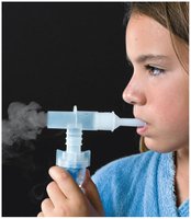
Cystic fibrosis is a genetic disorder characterized by the production of thick, sticky mucus that can obstruct airways and lead to recurrent respiratory infections.
Mucus accumulation blocks airflow and increases risk of bacterial infection.
Treatment often involves therapies to thin mucus and clear airways.
Anatomy of the Upper Respiratory System
Nose and Nasal Cavity
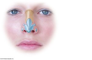
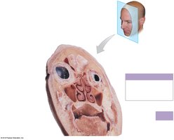
The nose is the primary entryway for air, which is filtered, warmed, and humidified as it passes through the nasal cavity. The nasal conchae increase surface area and turbulence, trapping particles in mucus.
Paranasal sinuses (maxillary, frontal, ethmoid, sphenoid) open into the nasal cavity and are lined with mucous membrane.
Tears draining through the nasal cavity help clean it.
Pharynx
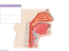
The pharynx is a muscular tube with three regions:
Nasopharynx: Functions in respiration only.
Oropharynx: Passageway for air and food.
Laryngopharynx: Passageway for air and food, leading to the larynx and esophagus.
Larynx and Voice Production
Structure of the Larynx
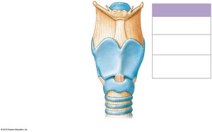
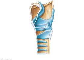
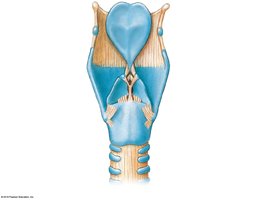
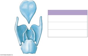
The larynx (voice box) connects the pharynx to the trachea and is involved in breathing, sound production, and protecting the lower airways.
Thyroid cartilage: Forms the Adam’s apple.
Epiglottis: Prevents food from entering the larynx during swallowing.
Cricoid cartilage: Connects the larynx to the trachea.
Voice Production
Sound is produced by the vibration of the vocal folds (true vocal cords) in the larynx. The pitch of the sound depends on the tension of the vocal folds, and articulation is achieved by the tongue, teeth, and lips.
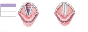

Lower Respiratory Tract: Trachea, Bronchi, and Bronchioles
Trachea
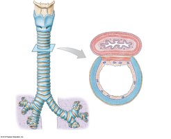
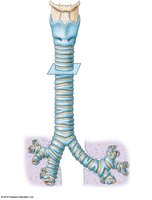
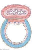
The trachea (windpipe) extends from the larynx to the primary bronchi. It is supported by C-shaped cartilage rings and lined with pseudostratified ciliated columnar epithelium.
Bronchi and Bronchioles
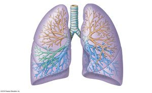
The trachea divides into right and left primary bronchi at vertebra T5. The right bronchus is wider and more vertical.
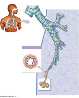
The bronchial tree branches into secondary (lobar) and tertiary (segmental) bronchi, then into bronchioles and terminal bronchioles.
Bronchioles have smooth muscle in their walls and are regulated by the autonomic nervous system (bronchodilation and bronchoconstriction).

Lungs and Pulmonary Anatomy
Gross Anatomy of the Lungs
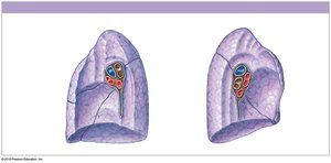
The lungs are paired organs protected by the pleural membranes (visceral and parietal pleura) with a lubricating pleural cavity between them.
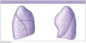
The right lung has three lobes (superior, middle, inferior) separated by horizontal and oblique fissures.
The left lung has two lobes (superior, inferior) and a cardiac notch.
Bronchopulmonary segments are supplied by tertiary bronchi and are surgically important subdivisions of the lung.
Pulmonary Lobules and Alveoli
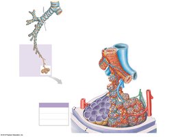
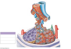
The smallest functional units of the lung are the pulmonary lobules, which contain alveoli where gas exchange occurs.
Air flows from terminal bronchioles to respiratory bronchioles, alveolar ducts, and alveoli.
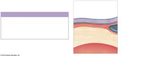
Alveoli are lined by type I pneumocytes (structural), type II pneumocytes (secrete surfactant), and alveolar macrophages (phagocytic).
Surfactant reduces surface tension, preventing alveolar collapse.
Respiratory Physiology
External and Internal Respiration
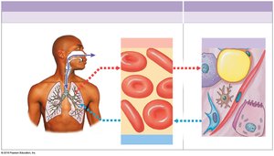
External respiration: Exchange of gases between alveoli and blood.
Internal respiration: Exchange of gases between blood and tissue cells.
Physical Principles of Ventilation
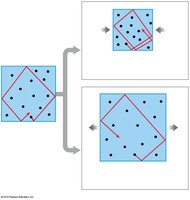
Air moves from areas of higher pressure to lower pressure (Boyle’s law: ).
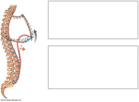
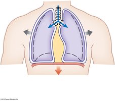
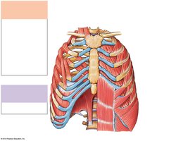
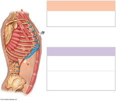
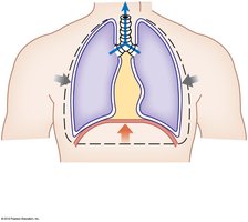
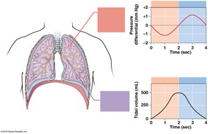
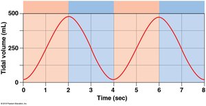
Inhalation occurs when thoracic volume increases (diaphragm and external intercostals contract), lowering intrapulmonary pressure.
Exhalation occurs when thoracic volume decreases (muscles relax or accessory muscles contract), raising intrapulmonary pressure.
Pulmonary Volumes and Capacities
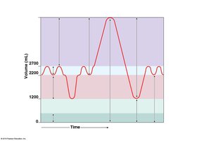
Tidal volume (TV): Air moved in and out during normal breathing (~500 mL).
Inspiratory reserve volume (IRV): Additional air inhaled after normal inspiration.
Expiratory reserve volume (ERV): Additional air exhaled after normal expiration.
Residual volume (RV): Air remaining after maximal exhalation.
Vital capacity (VC): IRV + TV + ERV.
Total lung capacity (TLC): VC + RV.
Gas Exchange and Transport
Gas Laws and Diffusion
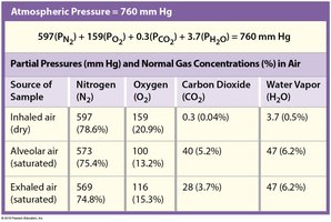
Dalton’s law: Each gas in a mixture exerts its own partial pressure; total pressure is the sum of all partial pressures.
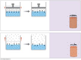
Henry’s law: The amount of gas dissolved in a liquid is proportional to its partial pressure and solubility.
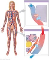
Oxygen Transport and Hemoglobin
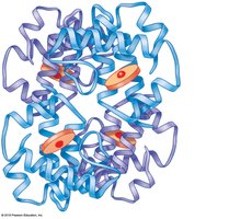
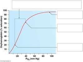
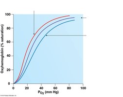
Most oxygen is transported bound to hemoglobin (Hb) as oxyhemoglobin (HbO2).
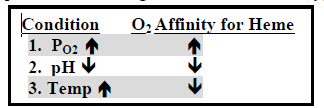
Hemoglobin’s affinity for O2 is affected by PO2, pH (Bohr effect), and temperature.
2,3-bisphosphoglycerate (BPG) produced by RBCs decreases Hb’s affinity for O2, promoting release in tissues.
Carbon Dioxide Transport
CO2 is transported dissolved in plasma, bound to hemoglobin (carbaminohemoglobin), and as bicarbonate ions (HCO3–).
High CO2 in blood promotes O2 release from Hb; high O2 promotes CO2 release from Hb.
Key reaction:
Clinical Considerations and Regulation
Pulmonary Disease: Compliance and Resistance
Compliance: Ease with which lungs expand. Decreased in fibrosis, increased in emphysema.
Resistance: Force required to move air. Increased in asthma, bronchitis, and with mucus or inflammation.
Neural Control of Respiration
Respiratory centers in the medulla oblongata and pons regulate the rate and depth of breathing.
Higher brain centers (cerebral cortex, hypothalamus, limbic system) can modify respiratory activity.
Reflex Control: Chemoreceptors and Baroreceptors
Chemoreceptors: Respond to changes in PCO2, PO2, and pH. Increased PCO2 is the strongest stimulus for increased ventilation.
Baroreceptors: Respond to changes in blood pressure, influencing respiratory rate.
Other reflexes include the inflation and deflation reflexes, sneezing, and coughing.
Effects of Aging and Smoking
Aging reduces all aspects of respiratory function.
Cigarette smoke contains carcinogens that can cause progressive changes in the respiratory epithelium, leading to cancer.
Progression: Dysplasia (reversible) → Metaplasia (reversible) → Neoplasia (irreversible) → Anaplasia (malignant, metastatic).
Medical Terminology
Asphyxia: Oxygen starvation due to low atmospheric oxygen or interference with ventilation or respiration.
Aspiration: Inhalation of foreign substances into the bronchial tree.
Dyspnea: Painful or labored breathing.
Epistaxis: Nosebleed.
