 Back
The Respiratory System: Structure, Function, and Regulation
Study Guide - Smart Notes
 Tailored notes based on your materials, expanded with key definitions, examples, and context.
The Respiratory System
Overview and Functional Anatomy
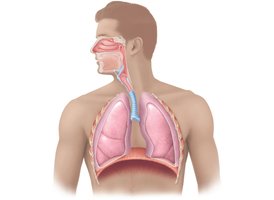
The respiratory system is responsible for the exchange of gases (oxygen and carbon dioxide) between the body and the environment. It consists of a series of organs and structures that facilitate breathing and gas exchange.
Major organs: Nose, nasal cavity, paranasal sinuses, pharynx, larynx, trachea, bronchi and their branches, lungs, and alveoli.
Primary functions: Air conduction, gas exchange, sound production, olfaction, and protection against pathogens.
Upper Respiratory System
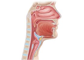
The upper respiratory tract includes the nose, nasal cavity, paranasal sinuses, and pharynx. These structures filter, warm, and moisten incoming air and serve as a passageway for airflow.
Nasal cavity: Lined with mucosa, contains nasal conchae to increase surface area and turbulence.
Pharynx: Divided into nasopharynx, oropharynx, and laryngopharynx; serves as a pathway for both air and food.
Larynx and Voice Production
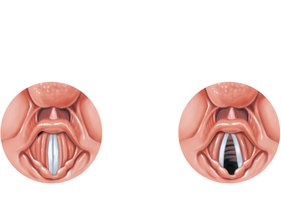
The larynx (voice box) is involved in sound production and protects the lower airways. The vocal folds (cords) vibrate to produce sound, with pitch and loudness determined by their length, tension, and the force of air.
Glottis: Opening between the vocal folds.
Pitch: Determined by the length and tension of the vocal cords.
Loudness: Depends on the force of air passing through the cords.
Lower Respiratory System
Conducting Zone Structures
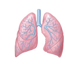
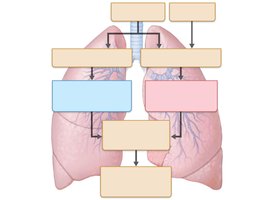
The conducting zone includes all respiratory passageways that conduct air to the respiratory zone. It consists of the trachea, bronchi, and bronchioles, which branch extensively within the lungs.
Trachea: Divides into right and left main (primary) bronchi.
Bronchi: Further branch into lobar (secondary) and segmental (tertiary) bronchi, then into bronchioles and terminal bronchioles.
Respiratory Zone Structures
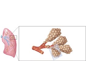
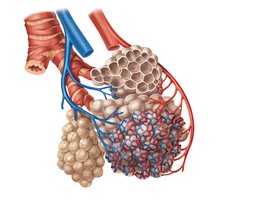
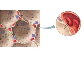
The respiratory zone is the site of gas exchange and includes respiratory bronchioles, alveolar ducts, and alveoli. Alveoli are tiny air sacs where oxygen and carbon dioxide are exchanged with the blood.
Alveoli: Approximately 300 million in the lungs, providing a large surface area for gas exchange.
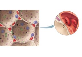
Respiratory membrane: Thin barrier formed by alveolar and capillary walls, facilitating efficient diffusion of gases.
Pleurae and Thoracic Cavity
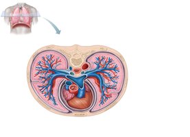
The lungs are enclosed by pleural membranes: the parietal pleura (lining the thoracic cavity) and the visceral pleura (covering the lungs). The pleural cavity contains fluid that reduces friction during breathing.
Mechanics of Breathing (Pulmonary Ventilation)
Phases of Breathing
Pulmonary ventilation consists of two phases: inspiration (air enters the lungs) and expiration (air exits the lungs). These processes depend on pressure changes within the thoracic cavity.
Inspiration: Active process involving contraction of the diaphragm and external intercostal muscles, increasing thoracic volume and decreasing intrapulmonary pressure.
Expiration: Usually passive; relaxation of inspiratory muscles decreases thoracic volume and increases intrapulmonary pressure, forcing air out.
Pressure Relationships
Atmospheric pressure (Patm): Pressure exerted by air around the body (760 mm Hg at sea level).
Intrapulmonary pressure (Ppul): Pressure within the alveoli; fluctuates with breathing but equalizes with Patm.
Intrapleural pressure (Pip): Pressure within the pleural cavity; always negative relative to Patm and Ppul.
Transpulmonary pressure: Difference between Ppul and Pip; keeps the lungs inflated.
Physical Factors Influencing Pulmonary Ventilation
Airway resistance: Usually low due to large airway diameters and extensive branching.
Alveolar surface tension: Reduced by surfactant (produced by type II alveolar cells), preventing alveolar collapse.
Lung compliance: Measure of lung expandability; decreased by fibrosis, reduced surfactant, or decreased thoracic flexibility.
Pulmonary Function Tests
Respiratory Volumes and Capacities
Pulmonary function tests assess lung function by measuring respiratory volumes and capacities using a spirometer.
Tidal volume (TV): Air inhaled or exhaled with each breath (about 500 ml in adults).
Inspiratory reserve volume (IRV): Additional air inhaled after a normal inspiration.
Expiratory reserve volume (ERV): Additional air exhaled after a normal expiration.
Residual volume (RV): Air remaining in lungs after forced expiration.
Vital capacity (VC): Maximum air exhaled after maximum inhalation:
Total lung capacity (TLC): Total air in lungs after maximum inspiration:
Dead Space
Anatomical dead space: Air in conducting zone not involved in gas exchange (~150 ml).
Alveolar dead space: Nonfunctional alveoli due to collapse or obstruction.
Total dead space: Sum of anatomical and alveolar dead space.
Gas Exchange
Composition of Alveolar Gas
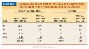
The composition of gases in the alveoli differs from atmospheric air due to gas exchange, humidification, and mixing of gases.
Gas | Atmosphere (%) | Atmosphere (mm Hg) | Alveoli (%) | Alveoli (mm Hg) |
|---|---|---|---|---|
N2 | 78.6 | 597 | 74.9 | 569 |
O2 | 20.9 | 159 | 13.7 | 104 |
CO2 | 0.04 | 0.3 | 5.2 | 40 |
H2O | 0.46 | 3.7 | 6.2 | 47 |
External and Internal Respiration
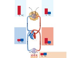
External respiration: Exchange of O2 and CO2 between alveoli and blood, driven by partial pressure gradients.
Internal respiration: Exchange of gases between blood and tissues; O2 diffuses into tissues, CO2 diffuses into blood.
Ventilation-Perfusion Coupling
Efficient gas exchange requires matching of ventilation (airflow) and perfusion (blood flow) in the lungs.
Control of Respiration
Neural Mechanisms
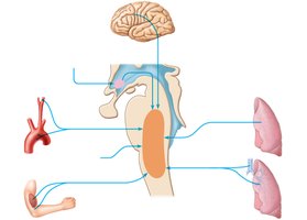
Breathing is controlled by respiratory centers in the medulla and pons. The ventral respiratory group (VRG) generates the basic rhythm, while the dorsal respiratory group (DRG) integrates sensory input and modifies the rhythm.
Normal rate: 12–15 breaths per minute in adults.
Factors Influencing Breathing Rate and Depth
Chemical factors: Levels of CO2, O2, and H+ in the blood are detected by chemoreceptors and influence respiratory rate and depth.
Other factors: Emotions, pain, body temperature, and pulmonary irritants can also affect breathing.
Respiratory Adjustments
Exercise: Increases ventilation (hyperpnea) to meet metabolic demands; blood gas levels remain relatively stable.
High altitude: Reduced atmospheric pressure can cause acute mountain sickness; long-term adaptation involves increased ventilation and red blood cell production.
Diving: Increased pressure affects lung volume and gas solubility; rapid ascent can cause barotrauma or decompression sickness ("the bends").
Homeostatic Imbalances of the Respiratory System
Chronic Obstructive Pulmonary Disease (COPD)
COPD is characterized by irreversible airflow limitation, commonly due to chronic bronchitis or emphysema. Most patients have a history of smoking.
Symptoms: Dyspnea, coughing, frequent infections, hypoxemia, and hypoventilation.
Pathology: Chronic inflammation, airway obstruction, destruction of alveolar walls, and loss of lung elasticity.
Asthma
Asthma is a reversible obstructive disease marked by airway inflammation, bronchospasm, and increased mucus production. It is often triggered by allergens or irritants and involves immune-mediated airway hyperresponsiveness.
Tuberculosis
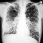
Tuberculosis is an infectious disease caused by Mycobacterium tuberculosis. It primarily affects the lungs and requires prolonged antibiotic treatment.
Symptoms: Fever, night sweats, weight loss, persistent cough, and hemoptysis (coughing up blood).
Lung Cancer
Lung cancer is the leading cause of cancer death in North America, with smoking as the primary risk factor. Major types include squamous cell carcinoma, adenocarcinoma, and small cell carcinoma.
Cystic Fibrosis
Cystic fibrosis is a genetic disorder caused by mutations in the CFTR gene, leading to thick, sticky mucus production, chronic respiratory infections, and progressive lung damage.
Additional info: The respiratory system is essential for maintaining homeostasis by regulating blood pH, facilitating speech, and protecting against environmental hazards.
