 Back
BackThe Respiratory System: Structure, Function, and Gas Exchange
Study Guide - Smart Notes
 Tailored notes based on your materials, expanded with key definitions, examples, and context.
The Respiratory System
Overview of Respiration
The respiratory system works closely with the circulatory system to supply oxygen (O2) to the body and remove carbon dioxide (CO2). Respiration involves four main processes: pulmonary ventilation, external respiration, transport of gases, and internal respiration.
Pulmonary ventilation: Movement of air into and out of the lungs.
External respiration: Exchange of O2 and CO2 between lungs and blood.
Transport: Movement of O2 and CO2 in the blood.
Internal respiration: Exchange of O2 and CO2 between systemic blood vessels and tissues.
Key Point: Blood going to tissues has higher O2 and lower CO2; blood returning from tissues has higher CO2 and lower O2.
Functional Anatomy of the Respiratory System
The respiratory system consists of several organs and structures that facilitate breathing and gas exchange.
Major organs: Nose, nasal cavity, paranasal sinuses, pharynx, larynx, trachea, bronchi, lungs, and alveoli.
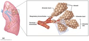
Respiratory zone: Site of gas exchange; includes respiratory bronchioles, alveolar ducts, and alveoli.
Conducting zone: Conduits to gas exchange sites; includes all other respiratory structures.
Respiratory muscles: Diaphragm and other muscles that promote ventilation.
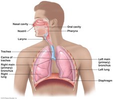
The Nose and Nasal Cavity
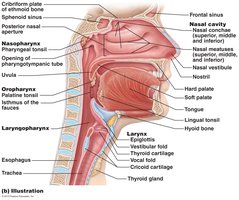
The nose provides an airway for respiration, moistens and warms incoming air, filters and cleans inspired air, serves as a resonating chamber for speech, and houses olfactory receptors.
Regions: External nose and nasal cavity.
Vestibule: Area superior to nostrils; vibrissae (hairs) filter coarse particles.
Olfactory mucosa: Contains smell receptors.
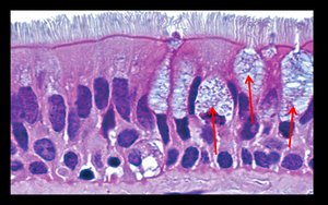
Respiratory mucosa: Pseudostratified ciliated columnar epithelium; contains lysozyme and defensins.
Cilia: Move contaminated mucus posteriorly to the throat.
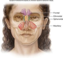
Nasal Conchae and Paranasal Sinuses
The nasal conchae are bony projections that increase mucosal area and enhance air turbulence, trapping particles. Paranasal sinuses lighten the skull and help warm and moisten air.
Conchae: Superior, middle, and inferior; protrude from lateral walls.
Sinuses: Located in frontal, sphenoid, ethmoid, and maxillary bones.
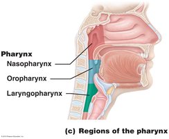
The Pharynx
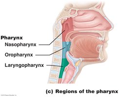
The pharynx is a muscular tube connecting the nasal cavity and mouth to the larynx and esophagus. It is divided into three regions: nasopharynx, oropharynx, and laryngopharynx.
Nasopharynx: Air passageway; lined with pseudostratified columnar epithelium.
Oropharynx: Passageway for food and air; lined with stratified squamous epithelium.
Laryngopharynx: Passageway for food and air; continuous with the esophagus.
The Larynx
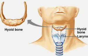
The larynx attaches to the hyoid bone and opens into the laryngopharynx, continuing with the trachea. It provides an open airway, routes air and food, and is involved in voice production.
Cartilages: Thyroid (Adam's apple), cricoid, arytenoid, cuneiform, corniculate, and epiglottis (elastic cartilage).
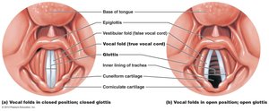
Vocal ligaments: Attach arytenoid to thyroid cartilage; form vocal folds (true vocal cords).
Glottis: Opening between vocal folds; vibrates to produce sound.
Vestibular folds: False vocal cords; help close glottis during swallowing.
The Trachea
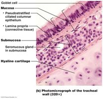
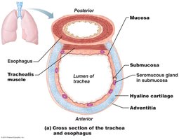
The trachea, or windpipe, extends from the larynx into the mediastinum. Its wall is composed of three layers: mucosa, submucosa, and adventitia, with C-shaped rings of hyaline cartilage.
Mucosa: Ciliated pseudostratified epithelium with goblet cells.
Submucosa: Connective tissue with seromucous glands.
Adventitia: Outermost connective tissue encasing cartilage rings.
Trachealis muscle: Connects posterior parts of cartilage rings; contracts during coughing.
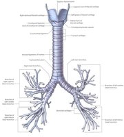
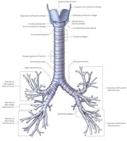
Carina: Last tracheal cartilage; point where trachea branches into bronchi.
Bronchi and Bronchial Tree
The bronchi undergo 23 orders of branching, forming the bronchial (respiratory) tree. The right main bronchus is wider, shorter, and more vertical than the left. Each main bronchus branches into lobar (secondary) bronchi, then segmental (tertiary) bronchi, bronchioles, and terminal bronchioles.
Structural changes: Cartilage rings give way to plates; absent in bronchioles. Epithelium changes from pseudostratified columnar to cuboidal; cilia and goblet cells become sparse. Smooth muscle increases.
Alveoli and Respiratory Membrane
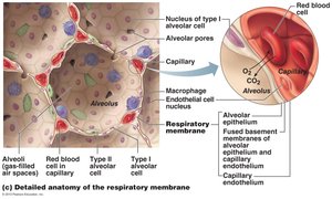
Alveoli are the main site for gas exchange, with about 300 million in the lungs. The respiratory membrane is a thin air-blood barrier consisting of alveolar and capillary walls and their fused basement membranes.
Alveolar walls: Single layer of squamous epithelium (type I cells); type II cells secrete surfactant and antimicrobial proteins.
Elastic fibers: Surround alveoli; open pores connect adjacent alveoli.
Alveolar macrophages: Keep alveolar surfaces sterile.
Lungs and Pleurae
The lungs occupy the thoracic cavity except the mediastinum. The left lung is smaller with two lobes; the right lung has three lobes. The pleurae are thin, double-layered serosae that provide lubrication and surface tension.
Root: Vascular and bronchial attachments.
Hilum: Site for attachment of blood vessels, bronchi, lymphatics, and nerves.
Pleurae: Parietal pleura on thoracic wall; visceral pleura on lung surface; pleural fluid fills pleural cavity.
Mechanics of Breathing
Pulmonary ventilation consists of inspiration (air into lungs) and expiration (air out of lungs). Volume changes in the thoracic cavity lead to pressure changes, causing gases to flow down their pressure gradient.
Boyle’s Law: (Pressure varies inversely with volume).
Inspiration: Active process; diaphragm and external intercostals contract, thoracic volume increases, intrapulmonary pressure drops, air flows in.
Expiration: Passive process; muscles relax, thoracic volume decreases, intrapulmonary pressure rises, air flows out.
Gas Exchange and Transport
Gas exchange occurs via external and internal respiration, governed by physical properties of gases and partial pressure gradients.
Dalton’s Law: Total pressure of a mixture of gases is the sum of the partial pressures of each gas.
Henry’s Law: Each gas dissolves in liquid in proportion to its partial pressure and solubility.
O2 transport: 98.5% bound to hemoglobin, 1.5% dissolved in plasma.
CO2 transport: 70% as bicarbonate ions, 20% bound to hemoglobin, 7-10% dissolved in plasma.
Regulation of Respiration
Respiratory control involves neurons in the medulla and pons, with chemical factors (CO2, O2, pH) influencing depth and rate of breathing.
Medullary respiratory centers: Dorsal and ventral groups regulate rhythm and integration.
Pontine centers: Smooth transitions between inspiration and expiration.
Chemoreceptors: Respond to changes in CO2, O2, and pH.
Homeostatic Imbalances
Several disorders affect the respiratory system, including hypoxia, chronic obstructive pulmonary disease (COPD), lung cancer, asthma, and tuberculosis.
Hypoxia: Inadequate O2 delivery to tissues.
COPD: Irreversible decrease in ability to force air out; includes chronic bronchitis and emphysema.
Lung cancer: Leading cause of cancer deaths; most cases due to smoking.
Asthma: Active inflammation of airways, bronchospasms, immune response.
Tuberculosis: Infectious disease caused by Mycobacterium tuberculosis.
