 Back
BackThe Respiratory System: Structure, Function, and Clinical Correlates
Study Guide - Smart Notes
 Tailored notes based on your materials, expanded with key definitions, examples, and context.
The Respiratory System
Anatomy of the Respiratory System: Overview
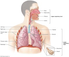
The respiratory system consists of organs and structures located in the head, neck, and thoracic cavity. It includes the pulmonary circuit blood vessels, thoracic cage structures, respiratory muscles, paired lungs, and the respiratory tract. The respiratory tract is divided into several regions:
Nose and Nasal Cavity – Located in cranial and facial bones
Pharynx (Throat)
Larynx – In the anterior neck
Trachea – In the mediastinum
Bronchial Tree – Begins as bronchi and divides into bronchioles
Divisions of the Respiratory Tract
Upper Respiratory Tract – Passageways from the nasal cavity to the larynx
Lower Respiratory Tract – Passageways from the trachea to the alveoli
Alveoli – Tiny air sacs for gas exchange; each lung contains about 150 million alveoli
Zones of the Respiratory System
Conducting Zone – Conduits for air movement (nose to bronchioles); air is filtered, warmed, and moistened
Respiratory Zone – Sites of gas exchange (structures containing alveoli only)
Basic Functions of the Respiratory System
Pulmonary Ventilation – Movement of air in and out of the lungs
Pulmonary Gas Exchange – Exchange of gases between lungs and blood
Gas Transport in the Blood – Movement of gases through the blood
Tissue Gas Exchange – Exchange of gases between blood and tissues
Other functions include speech production, odor detection, expulsion of abdominal contents, assisting venous and lymph flow, acid–base homeostasis, and blood pressure regulation.
Structures of the Upper Respiratory Tract
The Nose and Nasal Cavity
The nose and nasal cavity warm, humidify, and filter inhaled air, house olfactory receptors, and enhance voice resonance.
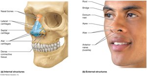
External Anatomy: Supported by nasal bones, lateral and alar cartilages; features include the root, bridge, dorsum nasi, apex, and alae (surrounding the nostrils).
Internal Anatomy: Hollow space divided by the nasal septum; contains nasal conchae (superior, middle, inferior) and meatuses, which create turbulent airflow for filtration and humidification.
Paranasal Sinuses: Frontal, ethmoid, sphenoid, and maxillary sinuses lighten the skull and enhance resonance.
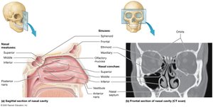
Histology: Vestibule lined with stratified squamous epithelium; olfactory mucosa on the roof; respiratory mucosa (pseudostratified ciliated columnar epithelium with goblet cells) lines the rest.
The Pharynx
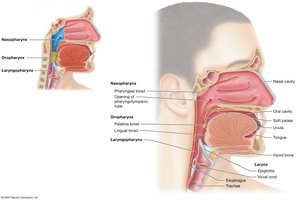
The pharynx is a muscular tube with three divisions:
Nasopharynx: Posterior to the nasal cavity; contains pharyngeal tonsil and pharyngotympanic tube openings; lined with pseudostratified ciliated columnar epithelium.
Oropharynx: Posterior to the oral cavity; contains palatine and lingual tonsils; lined with nonkeratinized stratified squamous epithelium.
Laryngopharynx: Extends from the hyoid bone to the esophagus; lined with nonkeratinized stratified squamous epithelium.
The Larynx
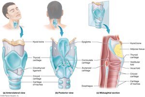
The larynx extends from the third to the sixth cervical vertebra and functions to keep food and liquids out of the lower respiratory tract and to house the vocal cords.
Cartilage Framework: Nine cartilages (thyroid, cricoid, epiglottis, arytenoid, corniculate, cuneiform); thyroid, cricoid, and most arytenoid are hyaline cartilage; epiglottis is elastic cartilage.
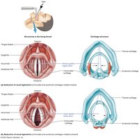
Vocal Folds: Vestibular folds (false vocal cords) and vocal folds (true vocal cords) produce sound; sound pitch and loudness depend on tension and force of air.
Lower Respiratory Tract
The Trachea
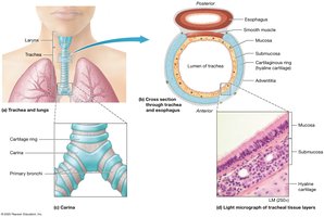
The trachea is a tube about 2 cm in diameter and 10–12 cm long, delivering air to the lower respiratory tract. It is supported by C-shaped rings of hyaline cartilage and lined with pseudostratified ciliated columnar epithelium.
Carina: Last tracheal cartilage ring; triggers cough reflex if foreign material is detected.
Layers: Mucosa (inner), submucosa (middle), adventitia (outer).
The Bronchial Tree
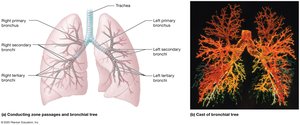
The bronchial tree consists of branching airways that conduct air to the alveoli:
Primary Bronchi: Right is wider, shorter, and straighter; left is narrower and more horizontal.
Secondary (Lobar) Bronchi: Three in the right lung, two in the left.
Tertiary (Segmental) Bronchi: About 10 per lung; further branching leads to bronchioles.
Bronchioles: Less than 1 mm in diameter, lack cartilage, have more smooth muscle, and simple cuboidal epithelium.
Terminal Bronchioles: Final part of conducting airways; branch into respiratory bronchioles and alveolar ducts.
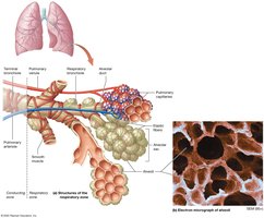
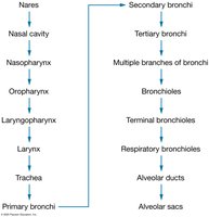
Alveoli and the Respiratory Membrane
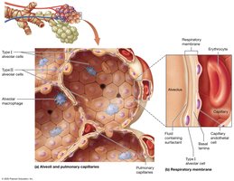
Alveoli are the primary sites of gas exchange and are surrounded by elastic fibers, smooth muscle, and pulmonary capillaries. Three main cell types are present:
Type I Alveolar Cells: Squamous cells for rapid gas diffusion (about 90% of alveolar wall).
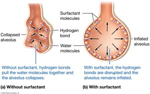
Type II Alveolar Cells: Cuboidal cells producing surfactant to reduce surface tension (about 10%).
Alveolar Macrophages: Phagocytes that remove debris.
The respiratory membrane consists of the alveolar epithelium, fused basal laminae, and capillary endothelium, and is extremely thin to facilitate gas exchange.
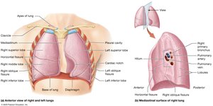
The Lungs and Pleurae
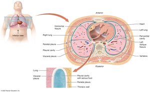
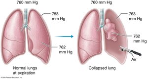
The lungs are divided into lobes (right: three; left: two) and bronchopulmonary segments. The pleural cavity is a double-layered serous membrane (parietal and visceral pleura) filled with pleural fluid for lubrication.
Mechanics of Breathing
Pressure-Volume Relationships
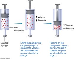
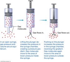
Breathing (pulmonary ventilation) depends on pressure gradients created by changes in lung volume. According to Boyle’s Law:
At constant temperature, pressure and volume are inversely related.
Phases of Pulmonary Ventilation
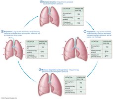
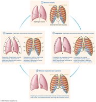
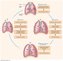
Inspiration: Diaphragm and external intercostals contract, increasing thoracic volume and decreasing intrapulmonary pressure, causing air to flow in.
Expiration: Muscles relax, thoracic volume decreases, intrapulmonary pressure increases, and air flows out.
Physical Factors Influencing Ventilation
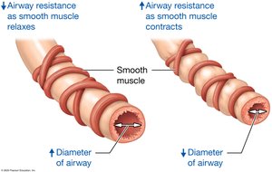
Airway Resistance: Increases with bronchoconstriction, decreases with bronchodilation.
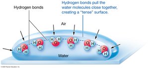
Alveolar Surface Tension: Reduced by surfactant, preventing alveolar collapse.
Pulmonary Compliance: Ability of lungs and chest wall to stretch; decreased by fibrosis or low surfactant.
Pulmonary Volumes and Capacities
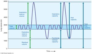
Pulmonary Volumes
Tidal Volume (TV): Air moved during normal breathing (~500 mL)
Inspiratory Reserve Volume (IRV): Additional air inspired after normal inspiration (2100–3300 mL)
Expiratory Reserve Volume (ERV): Additional air expired after normal expiration (700–1200 mL)
Residual Volume (RV): Air remaining after maximal expiration
Pulmonary Capacities
Inspiratory Capacity: TV + IRV
Functional Residual Capacity: ERV + RV
Vital Capacity: TV + IRV + ERV
Total Lung Capacity: TV + IRV + ERV + RV
Gas Exchange and Transport
Gas Laws
Dalton’s Law: Total pressure of a gas mixture is the sum of the partial pressures of each gas.
Henry’s Law: The amount of gas dissolved in a liquid is proportional to its partial pressure and solubility.
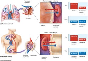
Pulmonary and Tissue Gas Exchange
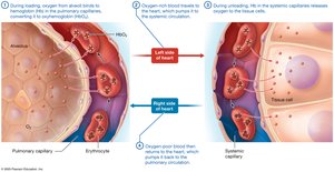
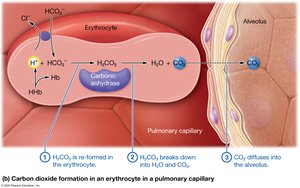
Pulmonary Gas Exchange: Oxygen moves from alveoli to blood; carbon dioxide moves from blood to alveoli.
Tissue Gas Exchange: Oxygen moves from blood to tissues; carbon dioxide moves from tissues to blood.
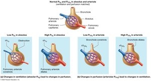
Ventilation–Perfusion Matching
Efficient gas exchange requires matching of air flow (ventilation) and blood flow (perfusion) in the lungs. Mismatches can lead to hypoxemia or alveolar dead space.

Oxygen and Carbon Dioxide Transport
Oxygen Transport
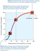
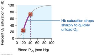
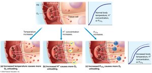
Hemoglobin (Hb): Each Hb molecule can bind four oxygen molecules; oxygen loading occurs in the lungs, unloading in the tissues.
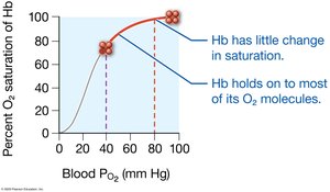
Oxygen–Hemoglobin Dissociation Curve: S-shaped curve showing the relationship between partial pressure of oxygen and Hb saturation.
Bohr Effect: Increased temperature, acidity, or 2,3-BPG decreases Hb affinity for oxygen, enhancing unloading in tissues.
Carbon Dioxide Transport
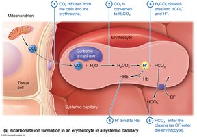
Dissolved in Plasma: 7–10%
Bound to Hemoglobin: 20% (as carbaminohemoglobin)
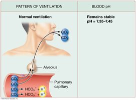
As Bicarbonate Ions: 70% (via carbonic anhydrase in erythrocytes)
Carbonic Acid–Bicarbonate Buffer System
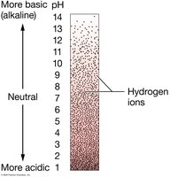
This system maintains blood pH within a narrow range (7.35–7.45):
Neural Control of Ventilation
Respiratory Centers
Respiratory Pattern Generator (RPG): Located in the medulla; sets basic rhythm of breathing.
Ventral Respiratory Group (VRG): Stimulates inspiratory muscles via phrenic and intercostal nerves.
Dorsal Respiratory Group (DRG): Integrates sensory information and relays it to other nuclei.
Chemoreceptor Regulation
Central Chemoreceptors: Detect changes in CO2 and pH in cerebrospinal fluid; increase ventilation if CO2 or H+ rises.
Peripheral Chemoreceptors: Located in carotid and aortic bodies; respond to low O2 or high H+ in arterial blood.
Clinical Correlates
Infectious Respiratory Diseases
COVID-19: Caused by SARS-CoV-2; symptoms range from mild to severe; can cause ARDS (acute respiratory distress syndrome).
Influenza: Caused by influenza viruses; symptoms include fever, cough, and fatigue; annual vaccination recommended.
Noninfectious Respiratory Diseases
Restrictive Lung Diseases: Decrease pulmonary compliance (e.g., idiopathic pulmonary fibrosis, pneumoconiosis).
Obstructive Lung Diseases: Increase airway resistance (e.g., COPD, emphysema, chronic bronchitis, asthma).
Lung Cancer: Tumors in the respiratory epithelium; leading cause of cancer death.
Additional info: This guide covers the structure and function of the respiratory system, mechanisms of breathing, gas exchange, transport, regulation, and common clinical conditions. It is suitable for college-level anatomy and physiology students preparing for exams.
