 Back
BackThe Respiratory System: Structure, Function, and Physiology
Study Guide - Smart Notes
 Tailored notes based on your materials, expanded with key definitions, examples, and context.
The Respiratory System: An Overview
Introduction to the Respiratory System
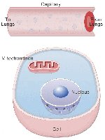
The respiratory system is essential for providing oxygen to body cells and removing carbon dioxide, a waste product of metabolism. This system works closely with the cardiovascular system to ensure efficient gas exchange and transport throughout the body.
Oxygen is required for ATP production in cells.
Carbon dioxide must be removed to prevent acid-base imbalance.
Gas exchange occurs via diffusion across lung surfaces.
Anatomy of the Respiratory System
Major Organs and Divisions
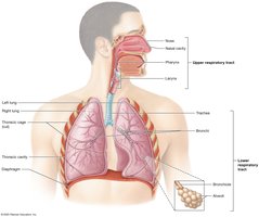
The respiratory system is divided into upper and lower regions, each with specific structures and functions.
Upper respiratory system: Nose, nasal cavity, paranasal sinuses, pharynx
Lower respiratory system: Larynx, trachea, bronchi, bronchioles, alveoli
Respiratory tract: Conducting portion (air movement) and respiratory portion (gas exchange)
Functions of the Respiratory System
Provides surface area for gas exchange
Moves air to and from exchange surfaces
Protects respiratory surfaces from pathogens and debris
Produces sounds (phonation)
Facilitates olfaction (smell)
Maintains acid-base balance
Histology of the Respiratory Tract
Respiratory Mucosa and Epithelium
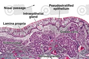
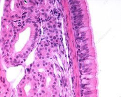
The respiratory mucosa lines the conducting portion of the tract and consists of an epithelial layer and a deep areolar layer (lamina propria). The type of epithelium changes along the tract:
Pseudostratified ciliated columnar epithelium with goblet cells: Nasal cavity, nasopharynx, superior lower respiratory tract
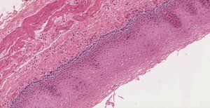
Stratified squamous epithelium: Inferior pharynx, oropharynx, laryngopharynx
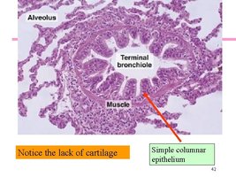
Cuboidal epithelium: Smaller bronchioles
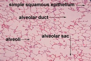
Simple squamous epithelium: Alveoli (site of gas exchange)
Respiratory Defense System
Filtration by nasal hairs removes large particles.
Mucous/goblet cells and glands produce mucus to trap debris.
Cilia sweep mucus toward the pharynx for expulsion or swallowing.
Alveolar macrophages engulf small particles in the lungs.
Upper Respiratory Tract
Nose and Nasal Cavity
The nose and nasal cavity filter, warm, and humidify incoming air. They are lined with pseudostratified ciliated columnar epithelium and contain bony projections called nasal conchae that create turbulence, enhancing filtration.
Pharynx
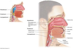
The pharynx is divided into three regions:
Nasopharynx: Pseudostratified ciliated columnar epithelium; contains pharyngeal tonsil
Oropharynx: Stratified squamous epithelium; contains palatine and lingual tonsils
Laryngopharynx: Stratified squamous epithelium
Larynx
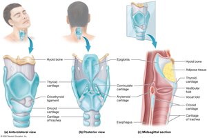
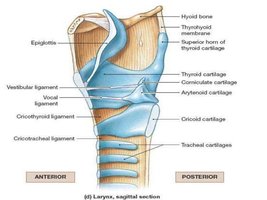
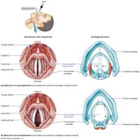
The larynx is a cartilaginous structure that protects the glottis and vocal cords. It is involved in sound production and prevents food from entering the lower respiratory tract during swallowing.
Main cartilages: Thyroid, cricoid, epiglottis
During swallowing, the epiglottis folds over the glottis
Lower Respiratory Tract
Trachea
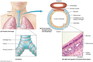
The trachea is supported by C-shaped rings of hyaline cartilage, keeping the airway open while allowing the esophagus to expand during swallowing. The carina at the base of the trachea triggers coughing if foreign material is detected.
Bronchial Tree
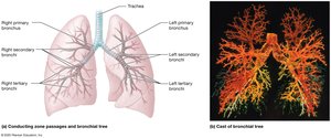
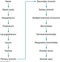
The bronchial tree consists of branching airways:
Primary bronchi → Secondary (lobar) bronchi → Tertiary (segmental) bronchi
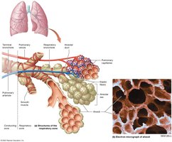
Bronchioles: Smallest airways, lack cartilage, lined by simple cuboidal epithelium
Terminal bronchioles: Final part of conducting airways
Alveoli and the Respiratory Membrane
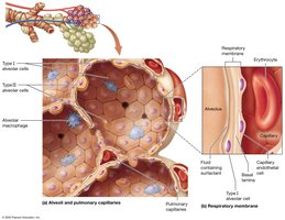
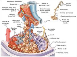
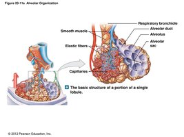
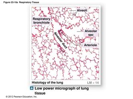
Alveoli are tiny air sacs where gas exchange occurs. The respiratory membrane consists of alveolar epithelium, capillary endothelium, and fused basement membranes, allowing efficient diffusion of gases.
Lungs and Pleurae
The lungs are divided into lobes and further into pulmonary lobules by connective tissue partitions. Each terminal bronchiole supplies a lobule, which contains respiratory bronchioles, alveolar ducts, and alveolar sacs. The lungs are surrounded by pleural membranes (parietal and visceral) with pleural fluid reducing friction during breathing.
Alveolar Epithelium and Surfactant
Type I alveolar cells: Simple squamous cells for gas diffusion
Type II alveolar cells: Produce surfactant, reducing surface tension and preventing alveolar collapse
Alveolar macrophages: Phagocytize debris and pathogens
Physiology of Respiration
Processes of Respiration
Pulmonary ventilation: Movement of air in and out of lungs
Pulmonary gas exchange: Exchange of gases between alveoli and blood
Gas transport: Movement of gases in the blood
Tissue gas exchange: Exchange of gases between blood and tissues
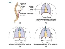
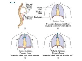
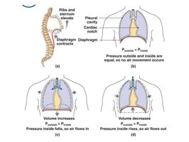
Pressure-Volume Relationships (Boyle's Law)
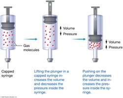
Boyle’s Law states that the pressure of a gas is inversely proportional to its volume, provided the number of gas molecules is constant:
Decreasing container size increases pressure
Increasing container size decreases pressure
Mathematically:
Mechanics of Breathing
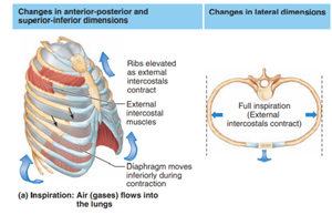
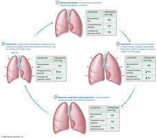
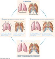
Breathing involves changes in thoracic cavity volume, creating pressure gradients that drive airflow:
Inhalation: Diaphragm contracts, ribcage elevates, thoracic volume increases, pressure decreases, air flows in
Exhalation: Diaphragm relaxes, ribcage lowers, thoracic volume decreases, pressure increases, air flows out
Respiratory Pressures
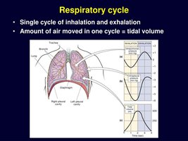
Intrapulmonary pressure: Pressure within alveoli, fluctuates with breathing
Intrapleural pressure: Pressure in pleural cavity, always less than atmospheric pressure
Tidal volume: Amount of air moved per breath
Respiratory Muscles
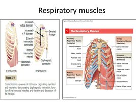
Diaphragm and external intercostals: Main muscles for quiet inhalation
Accessory muscles: Used during forced breathing
Normal exhalation is passive; forced exhalation uses accessory muscles
Physical Factors Influencing Ventilation
Airway resistance: Increases with constriction or obstruction
Alveolar surface tension: Reduced by surfactant to prevent collapse
Pulmonary compliance: Ability of lungs and chest wall to stretch
Gas Exchange and Transport
Pulmonary and Tissue Gas Exchange
Gas exchange occurs across the respiratory membrane in the lungs (external respiration) and between blood and tissues (internal respiration). Oxygen diffuses from alveoli to blood; carbon dioxide diffuses from blood to alveoli.
Oxygen Transport
Most oxygen is transported bound to hemoglobin in erythrocytes
Oxygen loading and unloading depend on partial pressures and affinity of hemoglobin
Carbon Dioxide Transport
Dissolved in plasma (7–10%)
Bound to hemoglobin as carbaminohemoglobin (20%)
As bicarbonate ions in plasma (70%)
Control of Respiration
Neural Control
Medullary respiratory centers: Dorsal (DRG) and ventral (VRG) respiratory groups
Pontine respiratory group: Modulates medullary centers
Regulate rate and depth of breathing based on blood gas levels
Chemoreceptor and Reflex Control
Central and peripheral chemoreceptors: Monitor CO2, O2, and pH
Stretch receptors: Respond to lung inflation
Irritant receptors: Trigger protective reflexes
Noninfectious Respiratory Diseases
Restrictive Lung Diseases
Decreased pulmonary compliance
Reduced inspiratory capacity, vital capacity, and total lung capacity
Obstructive Lung Diseases
Increased airway resistance, decreased efficiency of expiration
Examples: Chronic Obstructive Pulmonary Disease (COPD), emphysema, asthma
Asthma: Hyperresponsive airways, bronchoconstriction, inflammation, increased mucus
