 Back
BackThe Respiratory System: Structure, Function, and Clinical Aspects
Study Guide - Smart Notes
 Tailored notes based on your materials, expanded with key definitions, examples, and context.
CH 22 The Respiratory System: Overview and Functions
Major Functions of the Respiratory System
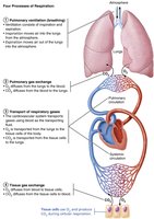
The respiratory system is essential for gas exchange, supplying oxygen to body cells and removing carbon dioxide produced during cellular respiration. It works closely with the cardiovascular system to ensure efficient transport and exchange of respiratory gases.
Pulmonary ventilation: Movement of air into and out of the lungs (breathing).
Pulmonary gas exchange: Exchange of gases (O2 and CO2) between the lungs and blood.
Transport of respiratory gases: Movement of gases in the blood between the lungs and tissues.
Tissue gas exchange: Exchange of gases between blood and body tissues.
Functional Anatomy of the Respiratory System
Major Organs and Divisions
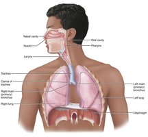
The respiratory system is divided into upper and lower regions, each with specialized structures for air conduction, filtration, and gas exchange.
Upper respiratory system: Nose, paranasal sinuses, pharynx
Lower respiratory system: Larynx, trachea, bronchi (and branches), lungs, alveoli
Respiratory muscles: Diaphragm and intercostal muscles (classified as part of the muscular system)
The Nose and Paranasal Sinuses
Structure and Functions
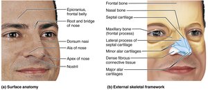
The nose is the only externally visible part of the respiratory system and serves as the main airway for respiration. It moistens, warms, filters, and cleans inspired air, acts as a resonating chamber for speech, and houses olfactory receptors.
External nose: Formed by nasal and frontal bones, maxillary bones, and hyaline cartilage.
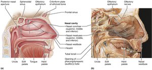
Nasal cavity: Divided by the nasal septum; lined with mucosa (olfactory and respiratory) and vibrissae (hairs) for filtration.
Nasal conchae: Increase surface area and enhance air turbulence for better filtration and humidification.
Paranasal sinuses: Lighten the skull, help warm and moisten air, and drain mucus into the nasal cavity.
The Pharynx
Regions and Functions
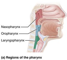
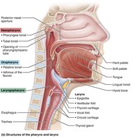
The pharynx (throat) is a muscular tube connecting the nasal cavity and mouth to the larynx and esophagus. It is divided into three regions:
Nasopharynx: Airway posterior to the nasal cavity; contains pharyngeal tonsils and auditory tubes.
Oropharynx: Passageway for food and air; contains palatine and lingual tonsils.
Laryngopharynx: Passageway for food and air; continuous with the esophagus.
The Larynx (Voice Box)
Structure and Function
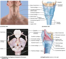
The larynx provides an open airway, routes air and food into proper channels, and houses the vocal folds for voice production. It is composed of nine cartilages, including the thyroid (Adam's apple), cricoid, arytenoid, cuneiform, corniculate, and the epiglottis (elastic cartilage).
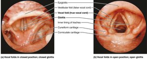
Vocal folds (true vocal cords): Vibrate to produce sound as air passes through.
Vestibular folds (false vocal cords): Help close the glottis during swallowing.
Epiglottis: Covers the laryngeal inlet during swallowing to prevent food entry into the airway.
The Trachea and Bronchial Tree
Trachea (Windpipe)
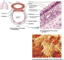
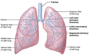
The trachea is a flexible tube supported by C-shaped cartilage rings, preventing collapse during breathing. It divides into right and left main bronchi, which enter the lungs at the hilum.
Wall layers: Mucosa (ciliated pseudostratified epithelium), submucosa (with seromucous glands), and adventitia (outer connective tissue).
Trachealis muscle: Allows constriction during coughing to expel mucus.
Bronchi and Subdivisions
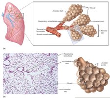
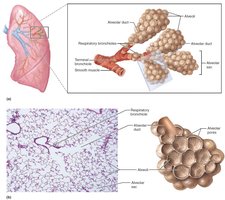
The bronchial tree undergoes about 23 generations of branching, forming conducting and respiratory zones. The conducting zone includes the trachea, bronchi, and bronchioles, while the respiratory zone includes respiratory bronchioles, alveolar ducts, and alveoli (sites of gas exchange).
The Lungs and Pleurae
Gross Anatomy of the Lungs
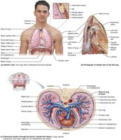
Each lung is divided into lobes (right: three; left: two) and further into bronchopulmonary segments. The lungs are soft, spongy, and highly elastic, surrounded by pleurae (serous membranes).
Pleurae: Parietal pleura lines the thoracic wall; visceral pleura covers the lungs. Pleural fluid lubricates and reduces friction during breathing.
Mechanics of Breathing
Pulmonary Ventilation
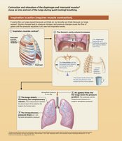
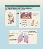
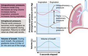
Breathing consists of inspiration (air in) and expiration (air out). Volume changes in the thoracic cavity lead to pressure changes, driving airflow.
Boyle’s Law: Pressure (P) of a gas varies inversely with its volume (V):
Inspiration: Diaphragm and external intercostals contract, increasing thoracic volume and decreasing pressure, drawing air in.
Expiration: Muscles relax, thoracic volume decreases, pressure increases, and air is expelled.
Pulmonary Volumes and Capacities
Measuring Lung Function
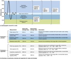
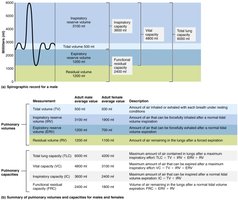
Pulmonary volumes and capacities are measured to assess respiratory health. Key volumes include:
Tidal volume (TV): Air moved per breath (~500 mL)
Inspiratory reserve volume (IRV): Extra air inhaled after normal inspiration
Expiratory reserve volume (ERV): Extra air exhaled after normal expiration
Residual volume (RV): Air remaining after forced expiration
Capacities are combinations of volumes (e.g., vital capacity, total lung capacity).
Gas Exchange and Transport
Gas Exchange Principles
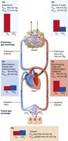
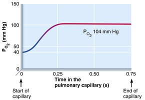
Gas exchange occurs by diffusion, driven by partial pressure gradients (Dalton’s Law) and solubility (Henry’s Law). Oxygen and carbon dioxide move between alveoli and blood, and between blood and tissues.
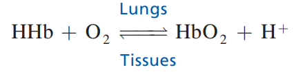
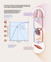
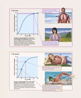
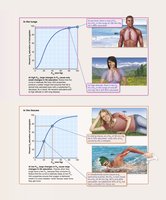
Oxygen transport: 98.5% bound to hemoglobin, 1.5% dissolved in plasma.
Carbon dioxide transport: Dissolved in plasma, bound to hemoglobin, or as bicarbonate ions.
Control of Respiration
Neural and Chemical Regulation
Respiratory rate and depth are controlled by centers in the medulla and pons, responding to chemical changes (CO2, O2, pH) detected by chemoreceptors.
Central chemoreceptors: Located in the brainstem; respond to CO2 and pH changes.
Peripheral chemoreceptors: Located in carotid and aortic bodies; respond to O2, CO2, and pH.
Clinical Aspects and Disorders
Common Respiratory Disorders
Rhinitis: Inflammation of nasal mucosa, often spreading to sinuses or lower respiratory tract.
Laryngitis: Inflammation of vocal folds, causing hoarseness or loss of voice.
Asthma: Reversible airway obstruction due to inflammation and bronchospasm.
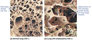
Chronic Obstructive Pulmonary Disease (COPD): Irreversible airflow limitation, including emphysema and chronic bronchitis.
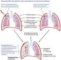
Pneumothorax: Air in the pleural cavity causing lung collapse.
Sleep apnea: Temporary cessation of breathing during sleep, often due to airway collapse.
Cystic fibrosis: Genetic disorder causing thick mucus and frequent lung infections.
Summary Table: Upper and Lower Respiratory Structures
Region | Main Structures | Functions |
|---|---|---|
Upper Respiratory | Nose, nasal cavity, paranasal sinuses, pharynx | Air conduction, filtration, humidification, olfaction |
Lower Respiratory | Larynx, trachea, bronchi, bronchioles, alveoli, lungs | Air conduction, voice production, gas exchange |
Key Equations
Boyle’s Law:
Oxygen-hemoglobin dissociation:
Additional info:
Clinical correlations and homeostatic imbalances are included to highlight the importance of respiratory health and common pathologies.
Tables and images are used to reinforce anatomical and physiological concepts.
