 Back
BackThe Respiratory System: Structure, Function, and Clinical Conditions
Study Guide - Smart Notes
 Tailored notes based on your materials, expanded with key definitions, examples, and context.
Respiratory System Overview
Introduction
The respiratory system is responsible for the exchange of gases (oxygen and carbon dioxide) between the body and the environment. It consists of a series of organs and structures that facilitate breathing, protect against pathogens, and contribute to vocalization.
Structural Plan of the Respiratory System
General Organization
The respiratory system resembles an inverted tree, with the trachea as the trunk and the alveoli as the leaves.
Gas exchange occurs by diffusion across the thin walls of the alveoli and capillaries.
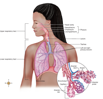
Respiratory Tracts
Upper and Lower Respiratory Tracts
Upper respiratory tract: Nose, pharynx, larynx
Lower respiratory tract: Trachea, bronchial tree, lungs
Head cold affects the upper tract (nose, sinuses, throat), while a chest cold affects the lower tract (trachea, bronchi, alveoli).
Respiratory Mucosa
Structure and Function
The respiratory mucosa is a mucous membrane lining the airways, producing over 125 mL of mucus daily.
Mucus traps dust and pathogens, while cilia move the mucus toward the pharynx for removal.
Respiratory membrane: The thin barrier between alveolar air and capillary blood, essential for gas exchange.
Nose and Paranasal Sinuses
Structure and Functions
The nasal septum divides the nose into two cavities, lined by mucous membrane.
Paranasal sinuses (frontal, maxillary, sphenoidal, ethmoidal) drain into the nose and contribute to voice resonance.
Olfactory receptors for smell are located in the nasal mucosa.
The nose warms, moistens, and filters inhaled air.
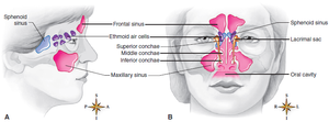
Pharynx (Throat)
Structure and Functions
The pharynx is about 12.5 cm long and divided into nasopharynx, oropharynx, and laryngopharynx.
It serves as a passageway for both air (respiratory system) and food (digestive system).
Tonsils (lymphoid tissue) provide immune protection but can cause breathing difficulty if swollen.
The eustachian tube connects the pharynx to the middle ear, equalizing pressure.
Tonsillitis
Clinical Condition
Tonsillitis is inflammation of the tonsils, often causing sore throat and difficulty swallowing.
Swollen tonsils can obstruct the airway, leading to dyspnea.
Larynx (Voice Box)
Structure and Functions
The larynx is located below the pharynx and is composed of several cartilages, including the thyroid cartilage (Adam’s apple).
The epiglottis covers the glottis during swallowing to prevent aspiration.
Vocal cords produce sound as air passes over them.
Trachea (Windpipe)
Structure and Function
The trachea is a tube about 11 cm long, supported by C-shaped cartilage rings to keep it open.
It is lined with ciliated mucosa that moves debris upward toward the pharynx.
Bronchi, Bronchioles, and Alveoli
Structure and Functions
The trachea divides into right and left primary bronchi, which branch into smaller bronchi and bronchioles.
Bronchioles end in clusters of alveolar sacs composed of alveoli, where gas exchange occurs.
Surfactant produced by type II alveolar cells reduces surface tension, preventing alveolar collapse.
Summary Table: Major Structures and Functions of the Respiratory System
Structure | Main Function(s) |
|---|---|
Nose | Warms, moistens, and filters air; sense of smell |
Pharynx | Passageway for air and food; immune defense (tonsils) |
Larynx | Voice production; air passage; prevents aspiration |
Trachea | Air passage; removal of debris by cilia |
Bronchi/Bronchioles | Air distribution to alveoli |
Alveoli | Gas exchange between air and blood |
Clinical Conditions of the Respiratory System
Upper Respiratory Infections (URIs)
Rhinitis: Inflammation of the nasal mucosa (common cold, allergies)
Pharyngitis: Sore throat due to infection or irritation
Laryngitis: Inflammation of the larynx, often causing hoarseness
Lower Respiratory Conditions
Acute bronchitis: Inflammation of the bronchi
Pneumonia: Infection causing fluid in alveoli
Tuberculosis: Chronic infection by Mycobacterium tuberculosis
COPD, asthma, emphysema: Chronic or obstructive disorders affecting airflow
Mechanics of Breathing
Pulmonary Ventilation
Inspiration: Active process; diaphragm and external intercostals contract, increasing thoracic volume and decreasing pressure, drawing air in.
Expiration: Usually passive; relaxation of inspiratory muscles and elastic recoil decrease thoracic volume, increasing pressure and expelling air.
Vital capacity (VC) is the maximum amount of air that can be exhaled after a maximal inhalation:
Where:
TV = Tidal Volume
IRV = Inspiratory Reserve Volume
ERV = Expiratory Reserve Volume
Regulation of Respiration
Control Centers and Receptors
Medullary rhythmicity area in the brainstem sets the basic rhythm of breathing.
Pontine centers adjust the rhythm as needed.
Chemoreceptors in the carotid and aortic bodies respond to changes in CO2, O2, and pH.
Pulmonary stretch receptors prevent overinflation of the lungs.
Gas Exchange and Transport
Mechanisms of Gas Exchange
Oxygen diffuses from alveoli into capillary blood, binding to hemoglobin to form oxyhemoglobin.
Carbon dioxide diffuses from blood into alveoli to be exhaled.
CO2 is transported in blood as dissolved gas (10%), carbaminohemoglobin (20%), and bicarbonate ions (70%).
This equation shows the conversion of carbon dioxide to bicarbonate in the blood, catalyzed by carbonic anhydrase.
Summary
The respiratory system is essential for gas exchange, vocalization, and protection against pathogens.
Its structure is adapted to maximize surface area for diffusion and to filter, warm, and humidify air.
Disorders can affect any part of the system, with symptoms and severity depending on the location and cause.
