 Back
BackThe Respiratory System: Structure, Function, and Clinical Implications
Study Guide - Smart Notes
 Tailored notes based on your materials, expanded with key definitions, examples, and context.
The Respiratory System
Overview and Primary Functions
The respiratory system is essential for gas exchange, supplying oxygen to body cells and removing carbon dioxide. It also plays roles in olfaction and speech. The respiratory and cardiovascular systems work together to ensure efficient transport and exchange of gases.
Pulmonary ventilation: Movement of air into and out of the lungs (breathing).
Pulmonary gas exchange: Exchange of O2 and CO2 between lungs and blood.
Tissue gas exchange: Exchange of O2 and CO2 between blood and tissues (handled by the cardiovascular system).
Protection: Respiratory surfaces are protected by mucous membranes, cilia, and immune cells that filter, moisten, and defend against pathogens and debris.
Organs of the Upper and Lower Respiratory Tract
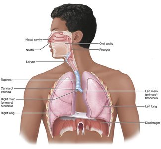
The respiratory tract is divided into upper and lower regions, each with specialized structures and functions.
Upper respiratory tract: Nose, paranasal sinuses, and pharynx.
Lower respiratory tract: Larynx, trachea, bronchi (and branches), and lungs (including alveoli).
Nose and Nasal Cavity
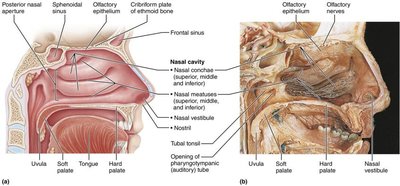
The nose is the only externally visible part of the respiratory system. It provides an airway, moistens and warms air, filters particles, serves as a resonating chamber for speech, and houses olfactory receptors. The nasal cavity is divided by the nasal septum and lined with mucosa that traps debris and pathogens.
Nasal vestibule: Lined with hairs (vibrissae) to filter large particles.
Olfactory mucosa: Contains olfactory epithelium for smell.
Respiratory mucosa: Contains cilia and mucus-secreting cells for trapping and moving debris.
Pharynx
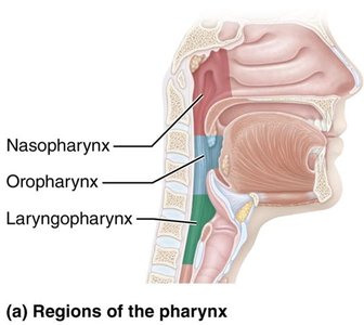
The pharynx (throat) connects the nasal cavity and mouth to the larynx and esophagus. It is divided into three regions:
Nasopharynx: Airway posterior to the nasal cavity.
Oropharynx: Passageway for food and air, posterior to the oral cavity.
Laryngopharynx: Passageway for food and air, continuous with the esophagus.
Larynx (Voice Box)
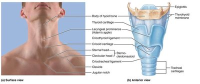
The larynx provides an open airway, routes air and food, and houses the vocal folds for sound production. It is composed of several cartilages, including the thyroid (Adam’s apple), cricoid, and epiglottis, which covers the laryngeal inlet during swallowing.
Trachea and Bronchial Tree
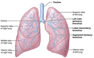
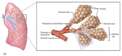
The trachea (windpipe) descends from the larynx and divides into right and left main bronchi. The bronchial tree undergoes multiple branchings, ending in bronchioles and terminal bronchioles. The walls contain cartilage for support and smooth muscle for regulating airflow.
Lungs and Alveoli
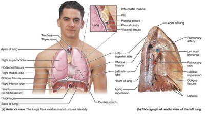
The lungs are divided into lobes and contain millions of alveoli, which are the primary sites of gas exchange. The respiratory membrane is extremely thin, allowing efficient diffusion of gases. Type II alveolar cells secrete surfactant to reduce surface tension, and macrophages keep the alveoli clean.
Pleurae
The lungs are surrounded by a double-layered pleura (parietal and visceral). The pleural cavity contains fluid to reduce friction during breathing. Inflammation of the pleura (pleurisy) or accumulation of fluid (pleural effusion) can impair breathing.
Processes of Respiration: External vs. Internal Respiration
Respiration involves several processes:
Pulmonary ventilation: Movement of air into and out of the lungs.
External respiration: Exchange of O2 and CO2 between alveoli and blood.
Internal respiration: Exchange of O2 and CO2 between blood and tissues.
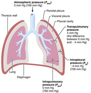
Pressure relationships in the thoracic cavity (atmospheric, intrapulmonary, and intrapleural pressures) drive ventilation. Boyle’s law describes the inverse relationship between pressure and volume in the lungs:
Clinical Implications: Atelectasis and Pneumothorax
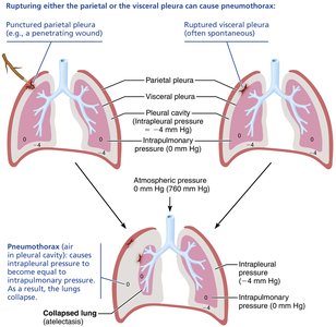
Atelectasis (lung collapse) can result from airway obstruction or air entering the pleural cavity (pneumothorax). Maintaining negative intrapleural pressure is essential for keeping the lungs inflated.
Physical Principles Governing Gas Diffusion
Gas exchange depends on partial pressure gradients, membrane thickness, and surface area. Pulmonary volumes (tidal volume, inspiratory/expiratory reserve, residual volume) are measured to assess lung function. Ventilation-perfusion coupling ensures efficient gas exchange by matching airflow and blood flow in the lungs.
Tidal volume (TV): Air moved per breath.
Inspiratory reserve volume (IRV): Extra air inhaled after a normal inspiration.
Expiratory reserve volume (ERV): Extra air exhaled after a normal expiration.
Residual volume (RV): Air remaining in lungs after forced expiration.
Hemoglobin and Gas Transport
Oxygen is transported in blood mainly bound to hemoglobin (Hb) in red blood cells. Each Hb molecule can bind four O2 molecules. The loading and unloading of O2 is influenced by partial pressure, temperature, pH, and CO2 levels. Carbon dioxide is transported dissolved in plasma, bound to Hb, or as bicarbonate ions.
Control of Respiration
Respiratory rate and depth are regulated by brainstem centers (medulla and pons), chemoreceptors (sensitive to CO2, O2, and pH), and reflexes. Hyperventilation can lead to decreased CO2 (hypocapnia), causing dizziness or fainting. The cyclic activity of inspiratory and expiratory neurons sets the normal breathing rhythm (eupnea).
Clinical Correlations
Rhinitis: Inflammation of the nasal mucosa, leading to congestion and postnasal drip.
Laryngitis: Inflammation of the vocal folds, causing hoarseness or loss of voice.
Pleurisy: Inflammation of the pleura, causing pain and impaired breathing.
Infant Respiratory Distress Syndrome (IRDS): Due to lack of surfactant in premature infants, leading to alveolar collapse.
Emphysema: Destruction of alveolar walls, reducing surface area for gas exchange.
Carbon monoxide poisoning: CO binds to Hb with high affinity, preventing O2 transport.
