 Back
BackThe Respiratory System: Structure, Function, and Mechanics of Breathing 23-1
Study Guide - Smart Notes
 Tailored notes based on your materials, expanded with key definitions, examples, and context.
The Respiratory System
Introduction to the Respiratory System
The respiratory system is essential for gas exchange, supplying oxygen (O2) to body cells for aerobic ATP production and removing carbon dioxide (CO2), a metabolic waste product. It consists of passageways in the head, neck, trunk, and the lungs, and is organized both structurally and functionally.
Passageway for air: Air moves between the atmosphere and alveoli during breathing.
Site for gas exchange: O2 diffuses from alveoli into blood; CO2 diffuses from blood into alveoli.
Olfaction: Olfactory receptors in the superior nasal cavity detect odors.
Sound production: Air movement across the vocal cords produces sound.
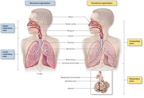
Structural and Functional Organization
Upper respiratory tract: Nose, nasal cavity, pharynx, larynx
Lower respiratory tract: Trachea, bronchi, bronchioles, alveolar ducts, alveoli
Conducting zone: Nose to terminal bronchioles (air transport only)
Respiratory zone: Respiratory bronchioles, alveolar ducts, alveoli (site of gas exchange)
Overview of Respiration
Processes of Respiration
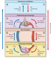
Respiration involves the movement of gases between the atmosphere and the body’s cells through four main processes:
Pulmonary ventilation: Movement of air between atmosphere and alveoli
Pulmonary gas exchange: Exchange of gases between alveoli and blood
Gas transport: Transport of gases in blood between lungs and systemic cells
Tissue gas exchange: Exchange of gases between blood and systemic cells
Anatomy of the Bronchial Tree
Bronchial Tree Structure
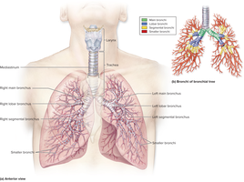
The bronchial tree is a system of highly branched air passages that originate at the main bronchi and branch into narrower tubes, ending in bronchioles.
Main bronchi (primary): Trachea splits into right and left main bronchi, each entering a lung.
Lobar bronchi (secondary): Each main bronchus branches into lobar bronchi, one for each lung lobe.
Segmental bronchi (tertiary): Further division into smaller passageways.
Bronchioles: Tubes <1 mm in diameter, leading to terminal bronchioles (end of conducting zone) and respiratory bronchioles (start of respiratory zone).
Histology and Functional Changes in the Bronchial Tree
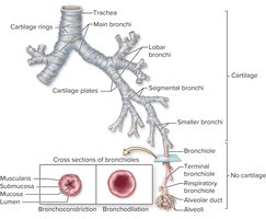
Main bronchi: Supported by incomplete rings of hyaline cartilage.
Bronchioles: No cartilage, but a thicker layer of smooth muscle.
Bronchoconstriction: Muscle contraction narrows bronchiole diameter, reducing airflow.
Bronchodilation: Muscle relaxation increases bronchiole diameter, increasing airflow.
Respiratory Zone: Bronchioles, Alveolar Ducts, and Alveoli
Microscopic Anatomy
The respiratory zone includes respiratory bronchioles, alveolar ducts, and alveoli, where gas exchange occurs.
Respiratory bronchioles: Lined with simple cuboidal epithelium.
Alveoli and alveolar ducts: Lined by simple squamous epithelium for efficient gas exchange.
Alveolar pores: Openings that provide collateral ventilation between alveoli.
Elastic fibers: Allow for expansion during inspiration and recoil during expiration.
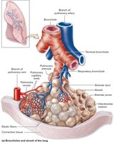
Cell Types of the Alveolar Wall
Alveolar type I cells: Squamous cells forming most of the alveolar surface and the respiratory membrane.
Alveolar type II cells (septal cells): Secrete pulmonary surfactant to reduce surface tension and prevent alveolar collapse.
Alveolar macrophages (dust cells): Engulf microorganisms and debris, either fixed in the wall or free to migrate.
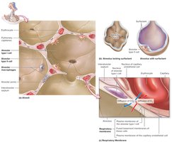
The Respiratory Membrane
Structure and Function
The respiratory membrane is a thin barrier (about 0.5 microns) that separates air in the alveoli from blood in pulmonary capillaries, facilitating rapid gas exchange.
Components: Plasma membrane of alveolar type I cell, plasma membrane of capillary endothelial cell, and their fused basement membranes.
Gas exchange: O2 diffuses from alveolus into capillaries; CO2 diffuses from blood to alveolus.
Pleural Membranes and Pressure Relationships
Pleural Structure and Pressure
Parietal pleura: Lines the thoracic wall.
Visceral pleura: Covers the lungs.
Pleural cavity: Space between pleurae containing pleural fluid.
Intrapleural pressure (Pip): Normally negative relative to intrapulmonary pressure, preventing lung collapse.
Transpulmonary pressure: Difference between intrapulmonary and intrapleural pressures; keeps lungs inflated.
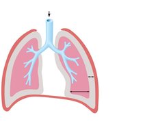
Pneumothorax
Pneumothorax is the presence of air in the pleural cavity, causing intrapleural pressure to equalize with atmospheric pressure, leading to lung collapse (atelectasis).
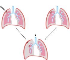
Mechanics of Breathing (Pulmonary Ventilation)
Muscles of Breathing
Breathing involves cyclic phases of inspiration (inhalation) and expiration (exhalation), regulated by skeletal muscles and neural control centers in the brain.
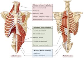
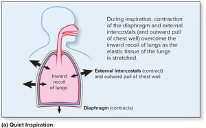
Quiet breathing: Diaphragm and external intercostals contract for inspiration; relax for expiration.
Forced breathing: Additional muscles contract for greater changes in thoracic volume and pressure.
Thoracic Cavity Volume Changes
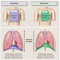
Vertical changes: Diaphragm contraction increases vertical dimension; relaxation decreases it.
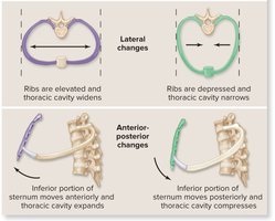
Lateral changes: Rib elevation widens, depression narrows the thoracic cavity.
Anterior-posterior changes: Sternum moves anteriorly in inspiration, posteriorly in expiration.
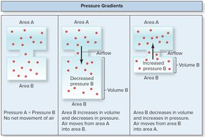
Boyle’s Law and Pressure Gradients
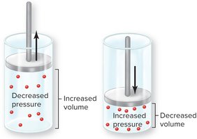
Boyle’s Law describes the inverse relationship between the pressure and volume of a gas at constant temperature:
As thoracic volume increases, intrapulmonary pressure decreases, causing air to flow into the lungs.
As thoracic volume decreases, intrapulmonary pressure increases, causing air to flow out.
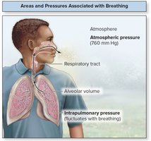
Volumes and Pressures Associated with Breathing
Atmospheric pressure: Pressure of air in the environment (760 mm Hg at sea level).
Intrapulmonary pressure: Pressure within alveoli; fluctuates with breathing.
Intrapleural pressure: Pressure within pleural cavity; always lower than intrapulmonary pressure.
Air flows down its pressure gradient (from high to low pressure).
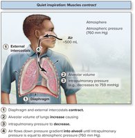
Mechanics of Quiet Breathing
Inspiration: Diaphragm and external intercostals contract, thoracic volume increases, intrapulmonary pressure drops below atmospheric pressure, air flows in (about 0.5 L as tidal volume).
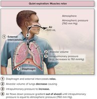
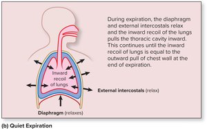
Expiration: Muscles relax, thoracic volume decreases, intrapulmonary pressure rises above atmospheric pressure, air flows out (about 0.5 L).
Clinical Views
Bronchitis
Acute bronchitis: Short-term inflammation, often following infection.
Chronic bronchitis: Long-term irritation, persistent cough, and mucus production; increases risk of infection.
Asthma
Episodes of bronchoconstriction, wheezing, coughing, and excess mucus due to localized immune reactions.
Treatment includes inhaled steroids and bronchodilators.
Pneumonia
Infection of the lung, causing alveoli to fill with fluid or pus, leading to impaired gas exchange.
Symptoms include cough, fever, difficulty breathing, and chest pain.
Summary Table: Key Structures and Functions of the Respiratory System
Structure | Main Function |
|---|---|
Nose/Nasal cavity | Warms, moistens, and filters air; olfaction |
Pharynx | Passageway for air and food |
Larynx | Voice production; airway protection |
Trachea | Conducts air to bronchi |
Bronchi/Bronchioles | Air passage; regulation of airflow |
Alveoli | Site of gas exchange |
