Back
BackThe Role of the CNS in Voluntary Movement
Study Guide - Smart Notes
 Tailored notes based on your materials, expanded with key definitions, examples, and context.
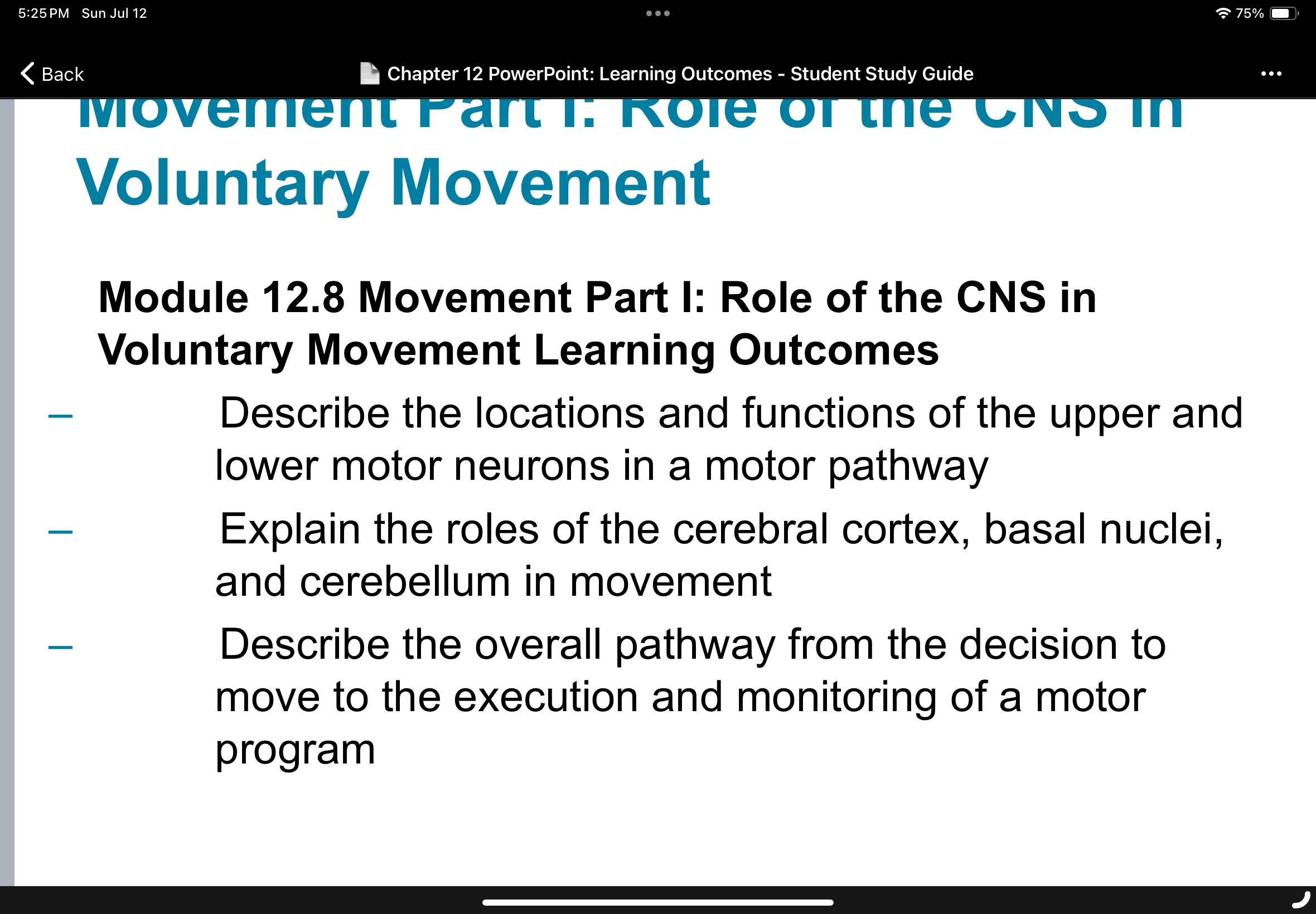
Module 12.8: Movement Part I – Role of the CNS in Voluntary Movement
Overview of Voluntary Movement
Voluntary movement is a complex process coordinated by multiple regions of the central nervous system (CNS). This module explores the neural pathways and structures involved in planning, initiating, executing, and monitoring voluntary motor actions.
Locations and Functions of Upper and Lower Motor Neurons
Motor pathways involve two main types of motor neurons: upper motor neurons and lower motor neurons.
Upper Motor Neurons: Located in the primary motor cortex of the cerebrum. Their axons descend through the brainstem and spinal cord, where they synapse with lower motor neurons. They are responsible for initiating and directing voluntary movements.
Lower Motor Neurons: Located in the anterior horn of the spinal cord or cranial nerve nuclei in the brainstem. Their axons exit the CNS to innervate skeletal muscles directly, causing muscle contraction.
Function: Upper motor neurons transmit signals from the brain to lower motor neurons, which then activate specific muscle fibers to produce movement.
Example: When deciding to pick up a cup, upper motor neurons in the motor cortex send signals down the spinal cord to lower motor neurons, which stimulate the muscles of the arm and hand.
Roles of the Cerebral Cortex, Basal Nuclei, and Cerebellum in Movement
Several CNS structures work together to ensure smooth, coordinated voluntary movements:
Cerebral Cortex: The primary motor cortex initiates voluntary movement. The premotor and supplementary motor areas are involved in planning and coordinating complex movements.
Basal Nuclei: These deep brain structures help regulate the initiation and termination of movements, suppress unwanted movements, and coordinate posture and muscle tone.
Cerebellum: The cerebellum monitors ongoing movements, compares intended movement with actual performance, and makes adjustments to ensure accuracy and coordination.
Example: The basal nuclei prevent involuntary tremors, while the cerebellum corrects errors during rapid hand movements.
Pathway from Decision to Move to Execution and Monitoring of a Motor Program
The process of voluntary movement involves several sequential steps:
Decision to Move: Initiated in the prefrontal cortex, where the intention to perform a movement is formed.
Motor Planning: The premotor cortex and supplementary motor area develop a motor program—a sequence of muscle contractions required for the movement.
Initiation: The primary motor cortex sends signals via upper motor neurons to the spinal cord.
Execution: Lower motor neurons activate the appropriate skeletal muscles.
Monitoring and Adjustment: Sensory feedback from muscles and joints is relayed to the cerebellum, which fine-tunes the movement as needed.
Example: When catching a ball, the brain plans the movement, initiates muscle contractions, and the cerebellum adjusts the arm's position in response to the ball's trajectory.