 Back
The Senses – Hearing: Anatomy and Physiology of the Ear
Study Guide - Smart Notes
 Tailored notes based on your materials, expanded with key definitions, examples, and context.
The Senses – Hearing
Introduction to Hearing
The ear is a complex sensory organ responsible for the special senses of hearing and equilibrium. This section focuses on the anatomical structures and physiological processes involved in hearing, from the external ear to the neural pathways that transmit sound information to the brain.
Anatomy of the Ear
External Ear
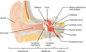
The external ear consists of the auricle (or pinna) and the auditory canal. The auricle is the visible, fleshy part of the ear that helps direct sound waves into the auditory canal. The canal leads to the tympanic membrane (eardrum), which marks the boundary between the external and middle ear.
Auricle (Pinna): Collects and directs sound waves toward the auditory canal.
Auditory Canal (External Acoustic Meatus): Channel through which sound waves travel to reach the tympanic membrane.
Tympanic Membrane: Vibrates in response to sound waves, transmitting vibrations to the middle ear.
Middle Ear
The middle ear is an air-filled cavity containing three small bones called auditory ossicles: the malleus, incus, and stapes. These bones transmit and amplify vibrations from the tympanic membrane to the oval window of the inner ear. The Eustachian tube connects the middle ear to the pharynx, equalizing air pressure on both sides of the tympanic membrane.
Malleus (Hammer): Attached to the tympanic membrane; transmits vibrations to the incus.
Incus (Anvil): Bridge between the malleus and stapes.
Stapes (Stirrup): Transmits vibrations to the oval window, initiating fluid movement in the inner ear.
Eustachian Tube: Equalizes pressure between the middle ear and the atmosphere.
Inner Ear
The inner ear is a bony labyrinth containing the cochlea (for hearing) and the vestibule (for balance). The cochlea is a spiral-shaped, fluid-filled structure that houses the sensory receptors for hearing. The vestibule and semicircular ducts are involved in equilibrium but are not discussed in detail here.
Cochlea: Contains the sensory organ for hearing (organ of Corti).
Vestibule: Involved in balance and equilibrium.
Oval Window: Membrane-covered opening that receives vibrations from the stapes.
Round Window: Allows for displacement of fluid within the cochlea.
Internal Structure of the Cochlea
Cochlear Ducts and the Spiral Organ
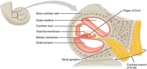
The cochlea contains three main chambers: the scala vestibuli, cochlear duct (scala media), and scala tympani. The cochlear duct houses the spiral organ (organ of Corti), which contains hair cells that act as mechanoreceptors for sound. The basilar membrane supports the spiral organ, while the tectorial membrane overlays the hair cells.
Scala Vestibuli: Upper chamber, begins at the oval window.
Cochlear Duct (Scala Media): Central chamber containing the spiral organ.
Scala Tympani: Lower chamber, ends at the round window.
Basilar Membrane: Supports the spiral organ and vibrates in response to sound-induced fluid waves.
Tectorial Membrane: Overlies the hair cells, facilitating their stimulation.
Hair Cells and Mechanotransduction
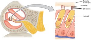
Hair cells within the spiral organ possess stereocilia that are deflected by movement of the basilar and tectorial membranes. This deflection opens ion channels, generating electrical signals that are transmitted to the brain via the cochlear branch of the vestibulocochlear nerve (CN VIII).
Hair Cells: Sensory receptors for hearing, located between the basilar and tectorial membranes.
Stereocilia: Hair-like projections that bend in response to fluid movement, initiating neural signals.
Sound Transduction Pathway
Pathway of Sound Waves
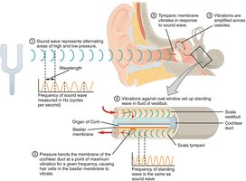
Sound waves are collected by the auricle, travel through the auditory canal, and vibrate the tympanic membrane. These vibrations are transmitted and amplified by the ossicles to the oval window, creating pressure waves in the cochlear fluids. The movement of these fluids causes the basilar membrane to vibrate, stimulating hair cells in specific regions according to sound frequency. The resulting neural signals are sent to the brain for interpretation as sound.
Sound waves enter the external ear and vibrate the tympanic membrane.
Vibrations are transmitted through the ossicles (malleus, incus, stapes).
The stapes moves the oval window, generating fluid waves in the cochlea.
Fluid waves move the basilar membrane, stimulating hair cells in the spiral organ.
Hair cells convert mechanical energy into electrical signals sent via the cochlear nerve to the brain.
Summary Table: Structures and Functions of the Ear
Structure | Function |
|---|---|
Pinna (Auricle) | Collects and directs sound waves into the auditory canal |
Auditory Canal | Conducts sound waves to the tympanic membrane |
Tympanic Membrane | Vibrates in response to sound waves; transmits vibrations to ossicles |
Malleus, Incus, Stapes | Transmit and amplify vibrations from tympanic membrane to oval window |
Oval Window | Receives vibrations from stapes; initiates fluid movement in cochlea |
Cochlea | Contains sensory receptors for hearing; transforms sound into neural signals |
Round Window | Allows for displacement of cochlear fluid |
Cochlear Nerve | Transmits auditory information to the brain |
Eustachian Tube | Equalizes air pressure across the tympanic membrane |
Additional Academic Context
Prolonged Exposure to Loud Noise: Chronic exposure to loud sounds can damage hair cells in the cochlea, leading to sensorineural hearing loss. Hair cells do not regenerate, making this damage permanent.
Frequency Coding: Different regions of the basilar membrane respond to different frequencies, allowing the ear to distinguish pitch.
Key Terms and Definitions
Audition: The sense of hearing.
Mechanoreceptor: A sensory receptor that responds to mechanical pressure or distortion.
Vestibulocochlear Nerve (CN VIII): The cranial nerve responsible for transmitting sound and equilibrium information from the inner ear to the brain.
