 Back
BackThe Sensory System: Structure and Function of Special Senses
Study Guide - Smart Notes
 Tailored notes based on your materials, expanded with key definitions, examples, and context.
Sensory System
Introduction to Special Senses
The sensory system is responsible for detecting changes in the environment and relaying this information to the nervous system for processing. Special senses include smell, taste, sight, hearing, and equilibrium. Each sense has specialized receptors and anatomical structures that allow for the detection and interpretation of specific stimuli.
The Eye and Vision
Anatomy of the Eye
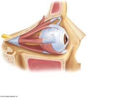
The eye is a complex organ containing over one million nerve fibers. It is protected and supported by several accessory structures:
Extrinsic eye muscles: Control eye movement.
Eyelids: Protect the eye and help spread tears.
Conjunctiva: Mucous membrane lining the eyelids and covering the sclera.
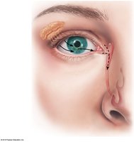
Lacrimal apparatus: Produces and drains tears, which cleanse and lubricate the eye.
External and Accessory Structures
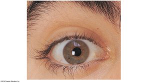
Eyelids: Meet at the medial and lateral commissures (canthi).
Eyelashes: Protect the eye; associated glands secrete lubricating fluids.
Lacrimal apparatus: Includes the lacrimal gland (produces tears) and ducts that drain into the nasal cavity.
Tears: Contain water, salts, mucus, antibodies, and lysozyme (an antibacterial enzyme).
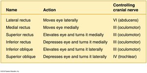
Extrinsic Eye Muscles
Six muscles attach to the outer surface of the eye, allowing for a wide range of movements.
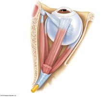
Name | Action | Controlling cranial nerve |
|---|---|---|
Lateral rectus | Moves eye laterally | VI (abducens) |
Medial rectus | Moves eye medially | III (oculomotor) |
Superior rectus | Elevates eye and turns it medially | III (oculomotor) |
Inferior rectus | Depresses eye and turns it medially | III (oculomotor) |
Inferior oblique | Elevates eye and turns it laterally | III (oculomotor) |
Superior oblique | Depresses eye and turns it laterally | IV (trochlear) |
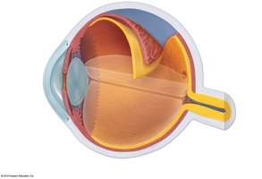
Internal Structures of the Eye
The eyeball is composed of three layers (tunics):
Fibrous layer: Sclera (white of the eye) and cornea (transparent front part).
Vascular layer: Choroid (blood-rich), ciliary body, and iris (controls pupil size).
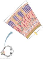
Sensory layer: Retina, containing photoreceptors (rods and cones).

Photoreceptors
Rods: Sensitive to dim light, provide black-and-white vision, concentrated at the retina's periphery.
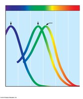
Cones: Responsible for color vision and visual acuity, concentrated in the fovea centralis.
Lens and Chambers
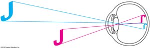
Lens: Flexible, biconvex structure that focuses light on the retina.
Anterior segment: Contains aqueous humor (maintains pressure, nourishes lens/cornea).
Posterior segment: Contains vitreous humor (maintains eye shape).
Physiology of Vision
Light is refracted by the cornea, aqueous humor, lens, and vitreous humor to focus on the retina.
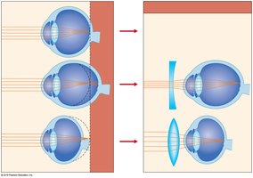
Accommodation: The lens changes shape to focus on near objects.
The image formed on the retina is real, inverted, and reversed left to right.
Visual Pathways
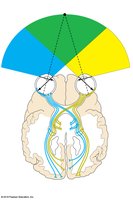
Impulses travel from the retina via the optic nerve, cross at the optic chiasma, continue through the optic tract, synapse in the thalamus, and reach the occipital lobe for interpretation.
Binocular vision provides depth perception.
Normal and Abnormal Vision
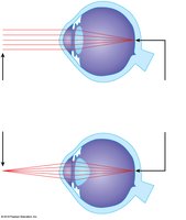
Emmetropia: Normal vision; image focused on the retina.
Myopia: Nearsightedness; image focused in front of the retina.
Hyperopia: Farsightedness; image focused behind the retina.
Astigmatism: Unequal curvature of cornea/lens causes blurred vision.
Eye Reflexes
Convergence: Eyes move medially for near vision.
Photopupillary reflex: Pupils constrict in bright light.
Accommodation pupillary reflex: Pupils constrict when focusing on close objects.
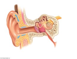
The Ear: Hearing and Balance
Anatomy of the Ear
The ear is divided into three regions:
External ear: Auricle (pinna) and external acoustic meatus (auditory canal).
Middle ear: Tympanic cavity, ossicles (malleus, incus, stapes), and pharyngotympanic tube.
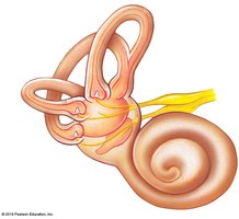
Internal ear: Bony labyrinth (cochlea, vestibule, semicircular canals) filled with perilymph and endolymph.
Equilibrium
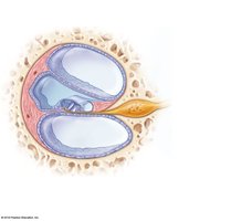
Vestibular apparatus: Contains receptors for static and dynamic equilibrium.
Static equilibrium: Maculae in the vestibule detect head position relative to gravity.
Dynamic equilibrium: Crista ampullaris in semicircular canals detects rotational movements.
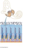
Process of Hearing
Sound waves are collected by the auricle, travel through the auditory canal, and vibrate the tympanic membrane.
Ossicles amplify vibrations and transmit them to the oval window of the cochlea.
Vibrations move the basilar membrane, bending hair cells in the spiral organ of Corti, generating nerve impulses.
Impulses travel via the cochlear nerve to the auditory cortex in the temporal lobe.
Hearing and Equilibrium Deficits
Conduction deafness: Impaired transmission of sound through the external/middle ear.
Sensorineural deafness: Damage to neural structures.
Ménière’s syndrome: Inner ear disorder causing deafness and vertigo.
Chemical Senses: Smell and Taste
Olfactory Receptors and the Sense of Smell
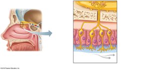
Olfactory receptors are located in the roof of the nasal cavity.
Chemicals must dissolve in mucus to be detected by olfactory hairs (cilia).
Impulses travel via the olfactory nerve (cranial nerve I) to the olfactory cortex for interpretation.
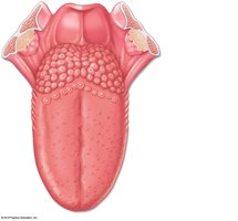
Taste Buds and the Sense of Taste
Taste buds are found on the tongue, soft palate, pharynx, and cheeks.
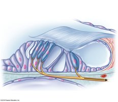
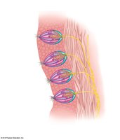
The tongue contains papillae: vallate (circumvallate), fungiform, and filiform.
Gustatory cells (taste receptors) have microvilli (gustatory hairs) that detect chemicals dissolved in saliva.
Impulses are carried by the facial (VII), glossopharyngeal (IX), and vagus (X) nerves.
Five basic tastes: sweet, sour, bitter, salty, umami.
Developmental Aspects of the Special Senses
Special sense organs develop early in embryonic life; maternal infections can cause abnormalities.
Vision matures after birth; infants are initially farsighted and lack depth perception.
Presbyopia (age-related loss of near vision) and presbycusis (age-related hearing loss) are common in aging.
Taste and smell are most acute at birth and decline after age 40.
