 Back
BackThe Skeletal System: Axial Gross Anatomy
Study Guide - Smart Notes
 Tailored notes based on your materials, expanded with key definitions, examples, and context.
The Skeletal System: Axial Gross Anatomy
Overview of the Skeletal System
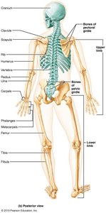
The human skeleton is divided into two main divisions: the axial skeleton and the appendicular skeleton. The axial skeleton forms the central axis of the body and includes the skull, vertebral column, and thoracic cage. The appendicular skeleton consists of the bones of the limbs and girdles that attach them to the axial skeleton.
Axial Skeleton: Skull, vertebral column, thoracic cage
Appendicular Skeleton: Limbs and girdles (pectoral and pelvic)
Bone Markings
Types and Functions of Bone Markings
Bone markings are specific features on bones that serve as sites for muscle, ligament, and tendon attachment, as well as passages for nerves and blood vessels, and points of articulation for joints.
Sites of muscle or ligament attachment: tuberosity, crest, trochanter, line, tubercle, epicondyle, spine
Sites involved in joints: head, condyle, ramus
Passages for nerves and blood vessels: meatus, sinus, fossa, foramen
Definitions:
Tuberosity: Large rounded projection; may be roughened
Crest: Narrow ridge of bone; usually prominent
Trochanter: Very large, blunt, irregularly shaped process (only on femur)
Line: Narrow ridge of bone; less prominent than a crest
Tubercle: Small rounded projection or process
Epicondyle: Raised area on or above a condyle
Spine: Sharp, slender, often pointed projection
Head: Bony expansion carried on a narrow neck
Condyle: Rounded articular projection
Ramus: Armlike bar of bone
Meatus: Canal-like passageway
Sinus: Cavity within a bone, filled with air and lined with mucous membrane
Fossa: Shallow, basinlike depression in a bone
Foramen: Round or oval opening through a bone
Bones of the Axial Skeleton
Major Components
The axial skeleton consists of the skull, vertebral column, and thoracic cage. These structures protect vital organs and provide support for the body.
Skull: Cranium and facial bones
Vertebral Column: Cervical, thoracic, lumbar vertebrae, sacrum, coccyx
Thoracic Cage: Sternum, ribs, and costal cartilages
The Skull
Cranium and Facial Bones
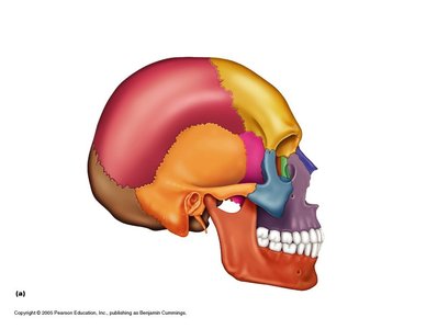
The skull is composed of two sets of bones: the cranium (which encloses the brain) and the facial bones (which form the structure of the face). The cranium consists of 8 bones, while the face is formed by 14 bones.
Cranial Bones (8): Frontal, Parietal (2), Occipital, Temporal (2), Sphenoid, Ethmoid
Facial Bones (14): Nasal (2), Maxillae (2), Zygomatic (2), Mandible, Lacrimal (2), Palatine (2), Inferior nasal conchae (2), Vomer
Major Sutures of the Skull
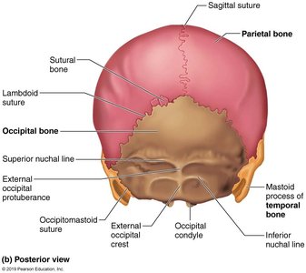
Sutures are immovable joints that connect the flat bones of the cranium. The four main sutures are:
Coronal suture: Between frontal and parietal bones
Sagittal suture: Between the two parietal bones
Squamous suture: Between temporal and parietal bones
Lambdoid suture: Between parietal and occipital bones
The Sphenoid Bone
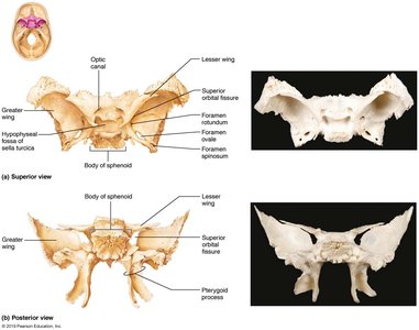
The sphenoid bone is a complex bone located at the base of the skull. It is considered the "keystone" of the cranial floor because it articulates with all other cranial bones.
Key features: Sella turcica, greater and lesser wings, pterygoid processes, optic canal, superior orbital fissure
The Ethmoid Bone
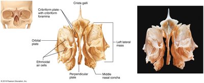
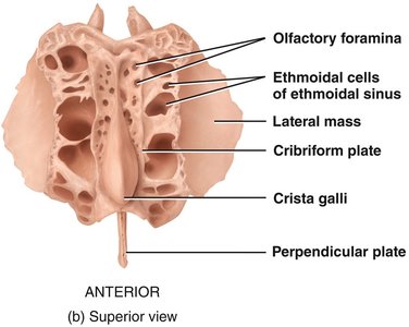
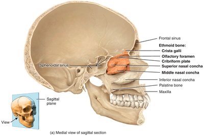
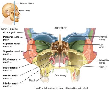
The ethmoid bone forms part of the anterior cranial fossa, the nasal septum, and the walls of the nasal cavity and orbits. It contains several important structures:
Cribriform plate: Contains olfactory foramina for passage of olfactory nerves
Crista galli: Attachment for the brain's dura mater
Perpendicular plate: Forms part of the nasal septum
Superior and middle nasal conchae: Increase surface area in the nasal cavity
Facial Bones and Markings
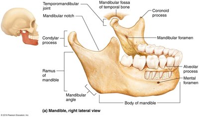
The facial bones provide the framework for the face, house the special sense organs, and provide openings for air and food passage. The mandible is the only moveable bone of the skull (excluding the ossicles of the middle ear).
Mandible: Body, ramus, coronoid process, condylar process, mandibular notch, alveolar process, mandibular foramen, mental foramen
Maxillae: Alveolar process, infraorbital foramen
Palatine: Horizontal plate, perpendicular plate
Bones of the Nasal Cavity and Paranasal Sinuses
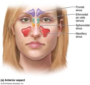
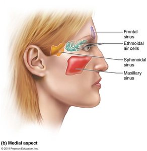
The nasal cavity is formed by several bones and contains structures that help warm, moisten, and filter air. The paranasal sinuses are air-filled spaces within certain skull bones that lighten the skull and enhance voice resonance.
Functions of nasal conchae: Increase surface area, warm and moisten air, trap particles
Paranasal sinuses: Located in the frontal, sphenoid, ethmoid, and maxillary bones
The Fetal Skull
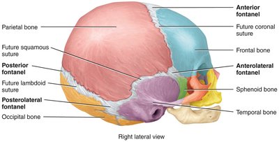
At birth, the bones of the skull are not fully ossified and are separated by fibrous membranes called fontanels. These allow for compression during childbirth and brain growth during infancy.
Fontanels: Anterior, posterior, sphenoidal (anterolateral), mastoid (posterolateral)
Closure: Fontanels close by about 2 years of age
Bone count: Babies have about 300 bones; adults have 206 due to fusion
The Vertebral Column
Regions and General Structure
The vertebral column, or spine, is composed of 26 bones in adults and provides the main support for the body while protecting the spinal cord. It is divided into five regions:
Cervical: 7 vertebrae (C1–C7)
Thoracic: 12 vertebrae (T1–T12)
Lumbar: 5 vertebrae (L1–L5)
Sacrum: 5 fused vertebrae
Coccyx: 3–5 fused vertebrae
Cervical Vertebrae (C1–C7)
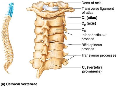
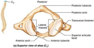
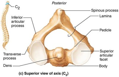
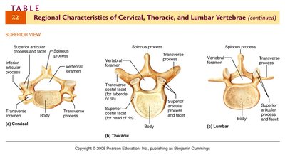
Cervical vertebrae are characterized by small bodies, bifid spinous processes, and transverse foramina. The first two cervical vertebrae, atlas (C1) and axis (C2), have unique features that allow for head movement.
Atlas (C1): No body or spinous process; articulates with occipital condyles for "yes" motion
Axis (C2): Has dens (odontoid process) for "no" motion
Thoracic Vertebrae (T1–T12)
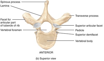
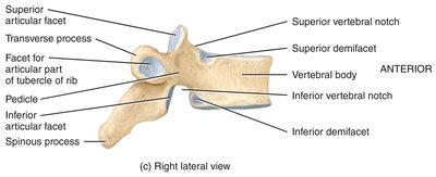
Thoracic vertebrae have heart-shaped bodies, long spinous processes, and facets for rib articulation. The vertebral foramen is circular.
Costal facets: For articulation with ribs
Lumbar Vertebrae (L1–L5)
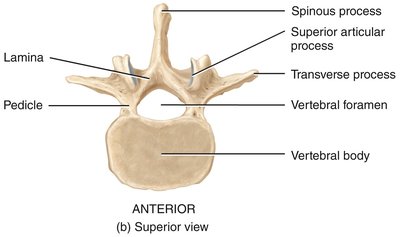
Lumbar vertebrae are the largest and strongest, with thick bodies for weight bearing and short, blunt spinous processes. The vertebral foramen is triangular.
Sacrum and Coccyx
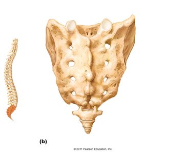
The sacrum is formed by the fusion of five vertebrae and articulates with the hip bones. The coccyx, or tailbone, is formed by the fusion of 3–5 small vertebrae and serves as an attachment site for ligaments and muscles.
Intervertebral Discs and Ligaments
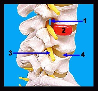
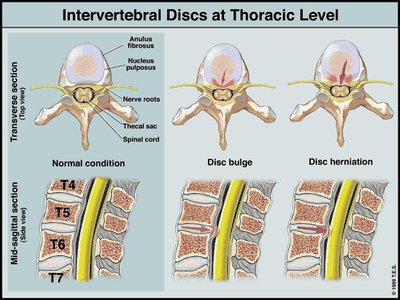
Intervertebral discs are composed of an outer ring of fibrocartilage (anulus fibrosus) and an inner gel-like core (nucleus pulposus). They absorb shock and allow flexibility. Ligaments and muscles provide additional support to the vertebral column.
Disorders of the Spine
Abnormal spinal curvatures can be congenital or acquired. Common disorders include:
Scoliosis: Abnormal lateral curvature
Kyphosis: Exaggerated thoracic curvature (hunchback)
Lordosis: Exaggerated lumbar curvature (swayback)
The Thoracic Cage (Bony Thorax)
Structure and Function
The thoracic cage protects vital organs in the thorax and supports the shoulder girdles and upper limbs. It consists of the sternum, ribs, thoracic vertebrae, and costal cartilages.
Sternum: Manubrium, body, xiphoid process
Ribs: 12 pairs; true ribs (1–7), false ribs (8–12), floating ribs (11–12)
Costal cartilage: Connects ribs to sternum
Table: Classification of Ribs
Type | Rib Numbers | Attachment |
|---|---|---|
True (vertebrosternal) | 1–7 | Directly to sternum via costal cartilage |
False (vertebrochondral) | 8–10 | Indirectly to sternum via cartilage of rib 7 |
Floating (vertebral) | 11–12 | No anterior attachment |
Example: The first rib is a true rib, directly attached to the sternum, while the eleventh rib is a floating rib with no anterior attachment.
Additional info: The thoracic cage also plays a role in respiration by allowing expansion and contraction of the lungs.
