 Back
BackThe Skeletal System: Bones, Cartilage, and Joints
Study Guide - Smart Notes
 Tailored notes based on your materials, expanded with key definitions, examples, and context.
The Skeletal System
Overview of the Skeletal System
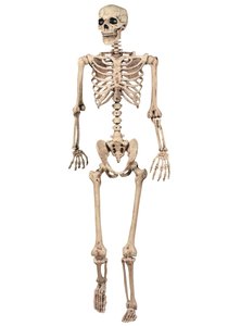
The skeletal system provides the structural framework for the human body, supporting movement, protecting organs, storing minerals, and housing bone marrow for blood cell formation. It is composed of bones and cartilage, which work together to maintain body shape and facilitate movement.
Support: Bones provide a rigid framework that supports the body and cradles soft organs.
Movement: Muscles attach to bones via tendons and use bones as levers to move the body.
Protection: Bones protect vital organs (e.g., skull protects the brain, rib cage protects the heart and lungs).
Mineral Storage: Bones store minerals such as calcium and phosphorus, releasing them into the bloodstream as needed.
Blood Cell Formation: Red bone marrow produces blood cells; yellow marrow stores fat.
Bone and Cartilage
Bones and cartilage are specialized connective tissues. In the developing embryo, the skeleton is initially composed mostly of cartilage, which is later replaced by bone during fetal and childhood development.
Bone: Hard, dense connective tissue that forms the majority of the adult skeleton.
Cartilage: Flexible connective tissue found in joints, ear, nose, and respiratory tract. It is more prevalent in embryos and children.
Types of Cartilage
Classification of Cartilage
Cartilage is classified into three main types based on the composition of their extracellular matrix and fibers:
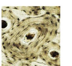
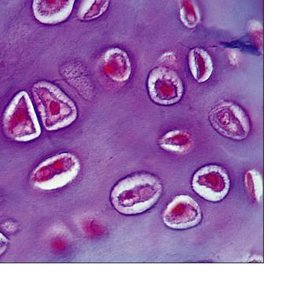
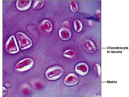
Hyaline Cartilage: Most common type; provides support with some flexibility. Found in the nose, trachea, larynx, and at the ends of long bones.
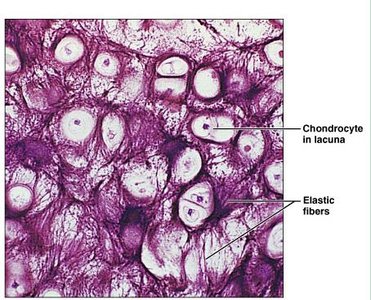
Elastic Cartilage: Contains elastic fibers, allowing for flexibility. Found in the external ear and epiglottis.
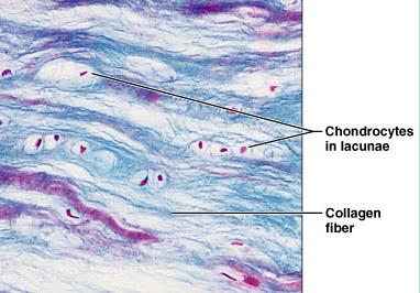
Fibrocartilage: Contains thick collagen fibers, providing strength and shock absorption. Found in intervertebral discs and pubic symphysis.
Classification of Bones
Bone Shapes
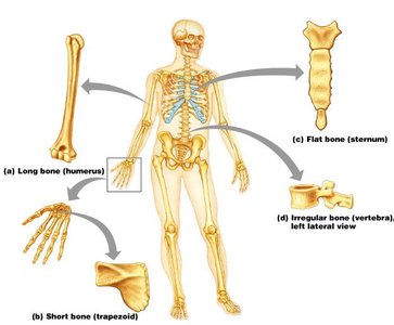
Bones are classified by their shapes, which relate to their functions and locations in the body:
Long Bones: Longer than they are wide (e.g., humerus, femur).
Short Bones: Nearly equal in length and width (e.g., carpals, tarsals).
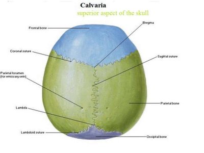
Flat Bones: Thin, flattened, and usually curved (e.g., sternum, ribs, skull bones).
Irregular Bones: Complex shapes (e.g., vertebrae, hip bones).
Pneumatic Bones: Contain air spaces (e.g., some skull bones).
Sesamoid Bones: Small, round bones embedded in tendons (e.g., patella).
Gross Anatomy of Bones
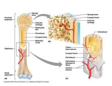
Bones have a complex structure with distinct regions and tissues:
Diaphysis: Shaft of a long bone.
Epiphysis: Ends of a long bone.
Metaphysis: Region between diaphysis and epiphysis; contains the growth plate in children.
Articular Cartilage: Hyaline cartilage covering joint surfaces.
Periosteum: Dense connective tissue covering the bone's outer surface.
Endosteum: Thin membrane lining the medullary cavity.
Medullary Cavity: Central cavity containing bone marrow.
Joints (Articulations)
Definition and Function
A joint, or articulation, is a site where two or more bones come together. Joints may permit movement, allow for growth, and transmit forces. They are classified by the range and type of movement they permit and by their anatomical structure.
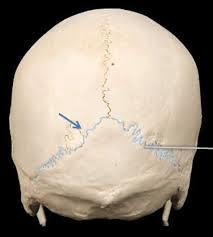
Immovable Joints (Synarthrosis): Permit little or no movement (e.g., sutures of the skull).
Slightly Movable Joints (Amphiarthrosis): Permit limited movement (e.g., intervertebral discs).
Freely Movable Joints (Diarthrosis): Permit a wide range of movements (e.g., shoulder, knee).
Structural Classification of Joints
Fibrous Joints: Bones connected by dense regular connective tissue; no joint cavity (e.g., sutures, syndesmoses, gomphoses).
Cartilaginous Joints: Bones united by cartilage; no joint cavity (e.g., synchondroses, symphyses).
Synovial Joints: Bones separated by a fluid-filled joint cavity; allow free movement.
Fibrous Joints
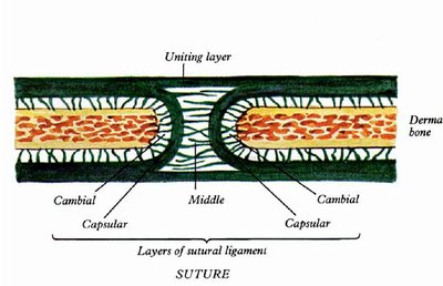
Sutures
Sutures are immovable joints found only between bones of the skull. The bones are joined by a thin layer of dense fibrous connective tissue, which is continuous with the periosteum. Sutures may ossify and fuse in middle age, forming synostoses.
Plane Suture: Simple apposition of flat bone surfaces.
Denticulate (Serrated) Suture: Interlocking, tooth-like projections (e.g., lambdoid suture).
Squamous Suture: Overlapping beveled edges (e.g., temporoparietal suture).
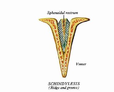
Schindylesis
Schindylesis is a type of fibrous joint where a ridge of one bone fits into a groove of another, such as between the vomer and the sphenoid bone.
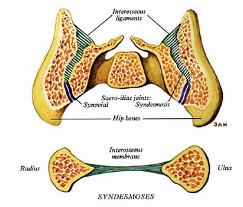
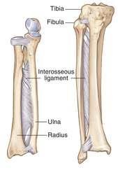
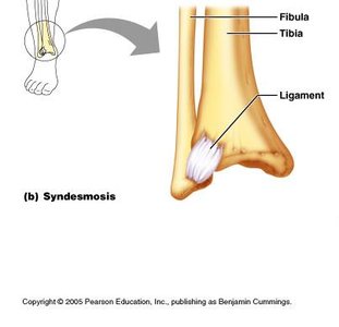
Syndesmosis
Syndesmoses are fibrous joints where bones are connected by ligaments or interosseous membranes. The amount of movement depends on the length of the connecting fibers.
Examples: Distal tibiofibular joint, interosseous membrane between radius and ulna.
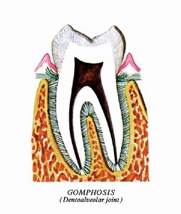
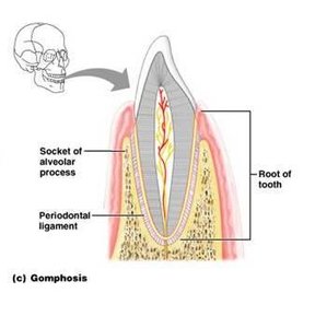
Gomphosis
Gomphosis is a peg-in-socket fibrous joint. The only example in the human body is the articulation of a tooth with its socket, held by the periodontal ligament.
Cartilaginous Joints
Synchondroses (Primary Cartilaginous Joints)
In synchondroses, bones are united by a plate or bar of hyaline cartilage. These joints are immovable and are found in the epiphyseal plates of growing bones and the joint between the first rib and the manubrium of the sternum.
Symphyses (Secondary Cartilaginous Joints)
Symphyses are slightly movable joints where bones are united by fibrocartilage. They provide strength, flexibility, and act as shock absorbers. Articular surfaces are covered by hyaline cartilage to reduce friction.
Examples: Intervertebral discs, pubic symphysis.
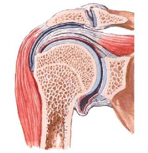
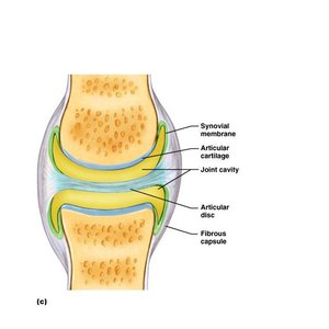
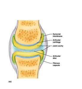
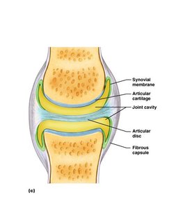
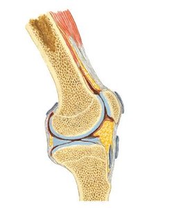
Synovial Joints (Diarthroses)
General Structure
Synovial joints are the most movable type of joint in the body. They are characterized by a fluid-filled joint cavity and a complex structure that allows for a wide range of movements.
Articular Cartilage: Hyaline cartilage covering bone ends, absorbing compression and protecting bones.
Joint (Synovial) Cavity: Space containing synovial fluid.
Articular Capsule: Two-layered capsule; outer fibrous layer and inner synovial membrane.
Synovial Fluid: Filtrate of blood, containing glycoproteins; lubricates and nourishes cartilage.
Reinforcing Ligaments: Capsular, extracapsular, and intracapsular ligaments strengthen the joint.
Nerves and Blood Vessels: Provide sensation and nutrients to the joint.
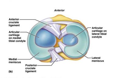
Articular Discs/Menisci: Pads of fibrocartilage that improve fit and absorb shock in some joints (e.g., knee, TMJ).
Fatty Pads: Cushioning structures found in some joints (e.g., hip, knee).
Bursae and Tendon Sheaths: Fluid-filled sacs that reduce friction between moving structures.
Classification of Synovial Joints by Movement
Uniaxial: Movement in one plane (e.g., hinge, pivot joints).
Biaxial: Movement in two planes (e.g., condylar, ellipsoid, saddle joints).
Multiaxial: Movement in multiple planes (e.g., ball and socket joints).
Non-axial: Sliding movements (e.g., plane joints).
Types of Synovial Joints
Plane Joints: Flat surfaces allow sliding movements (e.g., acromioclavicular joint).
Hinge Joints: Permit flexion and extension (e.g., elbow, knee, ankle).
Pivot Joints: Permit rotation around a single axis (e.g., atlantoaxial joint, radioulnar joint).
Condylar Joints: Permit flexion, extension, and limited rotation (e.g., knee, TMJ).
Ellipsoid Joints: Permit flexion, extension, abduction, adduction, and circumduction (e.g., wrist).
Saddle Joints: Permit flexion, extension, abduction, adduction, and rotation (e.g., thumb carpometacarpal joint).
Ball and Socket Joints: Permit movement in all axes (e.g., shoulder, hip).
Stability of Joints
The stability of a joint depends on the shape, size, and arrangement of articular surfaces, the strength of ligaments, and the tone of surrounding muscles.
Mechanism of Lubrication
Synovial fluid, hyaline cartilage, fatty pads, and bursae all contribute to reducing friction and facilitating smooth movement at synovial joints.
Nerve Supply of Joints
Joints receive a rich sensory nerve supply, which also innervates the muscles moving the joint and the overlying skin (Hilton's law).
Classification by Number of Bones
Simple Joint: Two bones (e.g., interphalangeal joint).
Compound Joint: More than two bones (e.g., elbow, wrist).
Complex Joint: Contains articular disc or meniscus (e.g., temporomandibular joint).
Key Movements at Joints
Flexion/Extension
Abduction/Adduction
Circumduction
Rotation
Examples and Applications
Lambdoid Suture: Example of a synarthrosis and fibrous joint.
First Sternocostal Articulation: Example of a synchondrosis.
Radioulnar Joint: Example of a pivot joint.
Symphysis Pubis: Example of a symphysis (cartilaginous joint).
Additional info: This guide covers the structure and classification of bones, cartilage, and joints, including their microscopic anatomy, functional roles, and clinical relevance for ANP college students.
