 Back
BackThe Skeletal System: Cartilage and Bone
Study Guide - Smart Notes
 Tailored notes based on your materials, expanded with key definitions, examples, and context.
The Skeletal System: Cartilage and Bone
Overview of the Skeletal System
The skeletal system is a complex organ system composed of bones, cartilage, nerves, blood vessels, and epithelial tissue. It provides structural support, protection, movement, mineral storage, and houses sites for blood cell formation and fat storage.
Bones: Hard, calcified matrix; adult human skeleton contains approximately 206 bones.
Cartilage: Provides flexibility and forms key structures in joints and other locations.
Other tissues: Nerves, blood vessels, and epithelial tissue are also present within the skeletal system.

Cartilage as a Connective Tissue
Cartilage is a specialized connective tissue that provides flexible support in various parts of the body. It is avascular (lacks blood vessels) and aneural (lacks nerves), which limits its ability to repair after injury.
Chondroblasts: Cells that secrete collagen and elastic fibers, forming the extracellular matrix.
Extracellular Matrix: Composed of about 80% water, dense irregular connective tissue, and fibers for support.
Perichondrium: Dense connective tissue membrane surrounding cartilage, containing blood vessels that nourish cartilage via diffusion.
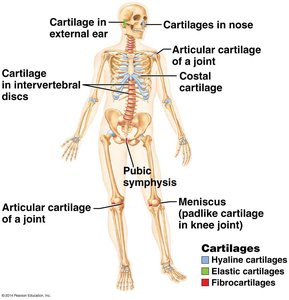
Types of Cartilage
There are three main types of cartilage, each with distinct characteristics and locations in the body:
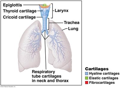
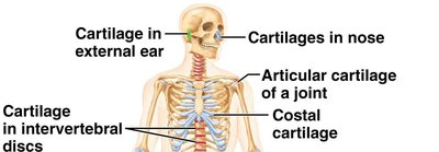
Hyaline Cartilage: Most abundant; provides firm support with some flexibility. Found in articular surfaces of joints, costal cartilages, nose, trachea, and larynx.
Elastic Cartilage: Contains more elastic fibers, allowing for repeated bending. Found in the epiglottis and external ear (pinna).
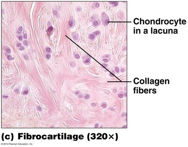
Fibrocartilage: Contains thick collagen fibers, making it strong and able to resist tension and absorb compressive forces. Found in intervertebral discs, menisci, and pubic symphysis.
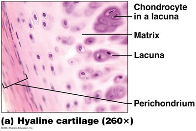
Hyaline Cartilage
Structure: Firm matrix with mostly collagen fibers; chondrocytes in lacunae.
Function: Cushions and resists compression.
Location: Articular cartilage of joints, costal cartilage, nose, trachea, larynx.
Elastic Cartilage
Structure: Similar to hyaline but with more elastic fibers.
Function: Maintains shape while allowing flexibility.
Location: Epiglottis, external ear (pinna).
Fibrocartilage
Structure: Thick collagen fibers; chondrocytes in lacunae.
Function: Resists tension and absorbs compressive forces.
Location: Intervertebral discs, menisci, pubic symphysis.
Functions of the Skeletal System
The skeletal system performs several essential functions for the human body:
Support: Provides a framework for the body and supports soft tissues.
Movement: Works with muscles to produce movement.
Protection: Protects vital organs such as the brain, spinal cord, and thoracic organs.
Mineral Reservoir: Stores calcium and phosphate ions.
Hemopoiesis: Houses red bone marrow for blood cell production.
Energy Storage: Stores fat in yellow bone marrow.
Endocrine Function: Osteoblasts secrete osteocalcin, a hormone involved in blood sugar regulation.
Types of Bone Tissue
Spongy Bone
Spongy bone, also known as cancellous bone, is composed of a network of trabeculae (rod-like structures) and is found primarily in the epiphyses of long bones and the interior of flat, short, or irregular bones.
Structure: Trabeculae lined with endosteum, containing osteocytes; spaces filled with red bone marrow.
Function: Resists forces from multiple directions and reduces skeletal weight.
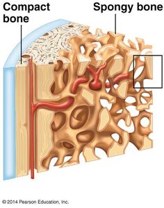
Compact Bone
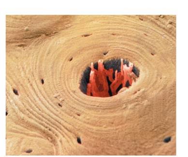
Compact bone forms the dense outer layer of bones and is organized into structural units called osteons (Haversian systems).
Osteons: Cylindrical structures running parallel to the long axis of the bone.
Lamellae: Concentric rings of bone matrix within osteons.
Lacunae: Small spaces between lamellae housing osteocytes.
Canaliculi: Tiny canals connecting lacunae, allowing nutrient and waste exchange between osteocytes.
Central (Haversian) Canal: Contains blood vessels and nerves.
Perforating (Volkmann's) Canals: Connect blood vessels of periosteum to those in central canals.
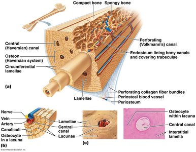
Microscopic Structure of Compact Bone
Interstitial Lamellae: Remnants of old osteons found between current osteons.
Circumferential Lamellae: Rings around the circumference of the diaphysis.
Collagen Fiber Orientation: Collagen fibers in adjacent lamellae run perpendicular, increasing bone strength.
Classification of Bones by Shape
Bones are classified based on their shapes, which relate to their functions:
Long Bones: Longer than they are wide (e.g., femur, humerus).
Short Bones: Nearly equal in length and width (e.g., carpals, tarsals).
Flat Bones: Thin, flattened, and often curved (e.g., sternum, ribs, skull bones).
Irregular Bones: Complex shapes (e.g., vertebrae, pelvic bones).
Anatomy of a Long Bone
Epiphyses: Ends of the bone, composed of spongy bone surrounded by compact bone; contains red bone marrow.
Diaphysis: Shaft of the bone; contains the medullary cavity (yellow marrow in adults, red marrow in children).
Epiphyseal Plate/Line: Growth plate (hyaline cartilage) in children; becomes the epiphyseal line (ossified) in adults.
Articular Cartilage: Hyaline cartilage covering joint surfaces.
Periosteum: Dense irregular connective tissue covering the outer surface (except at joints); contains osteoblasts and osteoclasts for bone remodeling.
Endosteum: Thin connective tissue lining internal bone surfaces, including trabeculae; also osteogenic.
Bone Growth and Development
Epiphyseal Plate vs. Epiphyseal Line
Epiphyseal Plate: Site of active growth in long bones; composed of hyaline cartilage.
Epiphyseal Line: Formed when the growth plate becomes ossified; indicates growth in length is complete.
Mechanisms of Bone Growth
Intramembranous Ossification: Bone develops from fibrous connective tissue membranes; forms most skull bones and clavicles.
Endochondral Ossification: Bone forms by replacing hyaline cartilage; forms most bones below the skull (except clavicles).
Bone Remodeling and Repair
Bone Remodeling
Bone remodeling is a continuous process involving osteoclasts (which break down bone matrix) and osteoblasts (which build new bone). Remodeling is regulated by mechanical stress and blood calcium levels.
Bone Fracture Healing
Formation of a hematoma (blood clot)
Formation of a fibrocartilaginous callus
Formation of a bony callus (spongy bone)
Bone remodeling restores original bone structure
Bone Disorders
Disorder | Mechanism | Common Causes | Effect on Bone | Strength |
|---|---|---|---|---|
Osteoporosis | Osteoclasts outpace osteoblasts | Low estrogen/testosterone, genetics, low calcium | Porous, fragile bone | Weak |
Osteomalacia (Rickets in children) | Insufficient mineralization of bone matrix | Vitamin D deficiency, poor calcium intake | Soft, flexible bones | Weak |
Paget's Disease | Excessive, disorganized bone remodeling | Hereditary/viral | Enlarged, deformed, less dense bones | Weak |
Role of Vitamin D in Bone Health
Vitamin D is synthesized in the skin upon exposure to UV light and is essential for calcium absorption in the digestive tract.
Calcium and phosphate ions are required for the formation of hydroxyapatite, the mineral component of bone.
Axial and Appendicular Skeleton
Axial Skeleton: 80 bones; includes the skull, vertebral column, and thoracic cage.
Appendicular Skeleton: 126 bones; includes the pectoral girdle, upper limbs, pelvic girdle, and lower limbs.
