Back
BackThe Skeletal System: Macro-to-Micro Blueprint (A&P Study Guide)
Study Guide - Smart Notes
 Tailored notes based on your materials, expanded with key definitions, examples, and context.
The Skeletal System: Macro-to-Micro Blueprint
Introduction to the Skeletal System
The skeletal system forms the rigid framework of the human body, providing structure, protection, and facilitating movement. It is a dynamic organ system, constantly remodeled and integrated with other physiological systems.

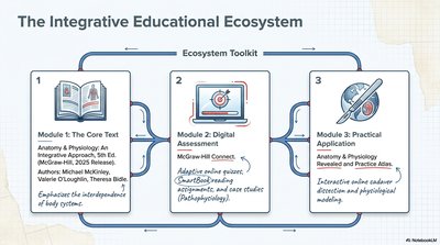
The Integrative Educational Ecosystem
Learning Modules for Anatomy & Physiology
Core Text: Comprehensive textbooks provide foundational knowledge and emphasize the interdependence of body systems.
Digital Assessment: Adaptive online quizzes and case studies reinforce learning and application.
Practical Application: Interactive atlases and digital models enhance understanding of structure and function.
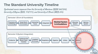
The Standard University Timeline
Course Sequence in Anatomy & Physiology
University-level A&P courses typically introduce the skeletal system after foundational topics such as cell biology, tissues, and the integumentary system. This sequencing allows for a comprehensive understanding of how the skeletal system integrates with other organ systems.
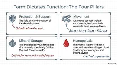
Form Dictates Function: The Four Pillars of the Skeletal System
Protection & Support: The skeleton provides a rigid framework that supports the body and protects internal organs.
Movement: Bones act as levers, joints as fulcrums, and muscles generate force for movement. Ligaments and tendons connect skeletal components.
Mineral Storage: Bones store essential minerals, primarily calcium (Ca) and phosphorus (P), critical for nerve and muscle function.
Hemopoiesis: The process of blood cell formation occurs in red bone marrow, producing erythrocytes, leukocytes, and thrombocytes.
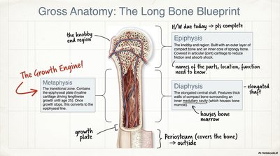
Gross Anatomy: The Long Bone Blueprint
Major Regions of a Long Bone
Epiphysis: The rounded end of the bone, covered with articular cartilage, involved in joint articulation.
Metaphysis: The growth region between the epiphysis and diaphysis, containing the epiphyseal plate (growth plate).
Diaphysis: The elongated shaft, housing the medullary cavity and bone marrow.
Periosteum: The outer fibrous covering of bone, essential for growth and repair.
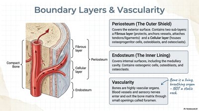
Boundary Layers & Vascularity
Bone Coverings and Blood Supply
Periosteum (Outer Shield): Composed of a fibrous layer (protection, attachment) and a cellular layer (osteogenic cells).
Endosteum (Inner Lining): Lines the medullary cavity, containing bone-forming and bone-resorbing cells.
Vascularity: Bones are highly vascularized, with blood vessels entering through foramina to nourish bone tissue.
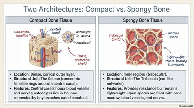
Two Architectures: Compact vs. Spongy Bone
Structural and Functional Differences
Compact Bone Tissue | Spongy Bone Tissue |
|---|---|
|
|
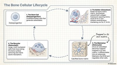
The Bone Cellular Lifecycle
Major Bone Cell Types and Functions
Osteoprogenitor Cells: Stem cells that differentiate into osteoblasts.
Osteoblasts (Builders): Synthesize bone matrix and initiate mineralization.
Osteocytes (Maintainers): Mature bone cells trapped in the matrix, regulate bone tissue.
Osteoclasts (Recyclers): Large, multinucleated cells that resorb bone matrix, essential for bone remodeling.
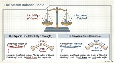
The Matrix Balance Scale
Organic vs. Inorganic Components of Bone
Organic Side (Flexibility & Strength): Composed mostly of osteoid (collagen). Provides tensile strength and flexibility.
Inorganic Side (Hardness): Composed of minerals, primarily calcium phosphate. Provides compressive strength and hardness.
Imbalances: Insufficient collagen leads to brittle bones; insufficient minerals lead to soft, bendable bones.
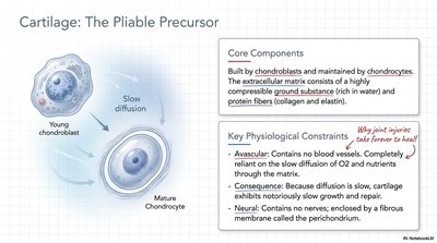
Cartilage: The Pliable Precursor
Structure and Function of Cartilage
Core Components: Built by chondroblasts and maintained by chondrocytes. The extracellular matrix is rich in water and protein fibers (collagen, elastin).
Physiological Constraints: Cartilage is avascular (no blood vessels), relies on slow diffusion for nutrients, and is enclosed by the perichondrium. It is also aneural (no nerves).
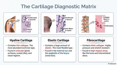
The Cartilage Diagnostic Matrix
Types of Cartilage and Their Locations
Hyaline Cartilage | Elastic Cartilage | Fibrocartilage |
|---|---|---|
|
|
|
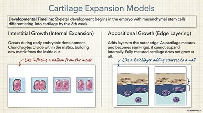
Cartilage Expansion Models
Growth Mechanisms of Cartilage
Interstitial Growth: Chondrocytes divide within the matrix, expanding cartilage from the inside (common in early development).
Appositional Growth: New layers are added to the surface by chondroblasts in the perichondrium (common in mature cartilage).
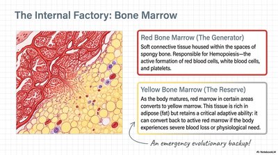
The Internal Factory: Bone Marrow
Types and Functions of Bone Marrow
Red Bone Marrow: Responsible for hemopoiesis (formation of blood cells). Found in spongy bone spaces.
Yellow Bone Marrow: Stores fat (adipose tissue) and can revert to red marrow under physiological stress.
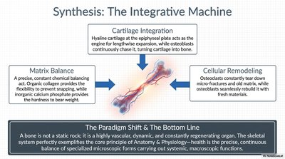
Synthesis: The Integrative Machine
Dynamic Balance and Remodeling in Bone
Cartilage Integration: Hyaline cartilage at the epiphyseal plate enables bone elongation, with osteoblasts converting cartilage to bone.
Matrix Balance: Organic collagen provides flexibility; inorganic calcium phosphate provides hardness.
Cellular Remodeling: Osteoclasts and osteoblasts continuously remodel bone, maintaining strength and adapting to stress.
Bottom Line: Bone is a highly vascular, dynamic, and constantly regenerating organ, exemplifying the integration of microscopic and macroscopic functions in anatomy and physiology.