 Back
BackThe Skeletal System: Structure and Organization
Study Guide - Smart Notes
 Tailored notes based on your materials, expanded with key definitions, examples, and context.
Skeletal System Organization
Overview of the Skeletal System
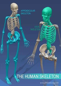
The human skeletal system is a complex framework composed of bones, cartilage, ligaments, and connective tissues. It provides structural support, protects vital organs, and serves as an attachment site for muscles, facilitating movement. The skeleton is divided into two main divisions: the axial skeleton and the appendicular skeleton.
Axial Skeleton: Forms the central axis of the body and includes the skull, vertebral column, and thoracic cage.
Appendicular Skeleton: Comprises the bones of the limbs and the girdles that attach them to the axial skeleton.
Axial vs. Appendicular Skeleton
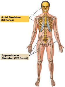
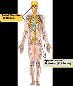
The axial skeleton consists of 80 bones, including the skull, vertebral column, and thoracic cage. It supports and protects the brain, spinal cord, and organs in the thorax, and provides attachment points for muscles involved in head, neck, and trunk movements. The appendicular skeleton contains 126 bones, including the limbs and the pectoral and pelvic girdles, which connect the limbs to the trunk and facilitate movement.
Axial Skeleton
Components of the Axial Skeleton
The axial skeleton forms the longitudinal axis of the body and is divided into three main parts:
Skull
Vertebral column
Bony thorax (thoracic cage)
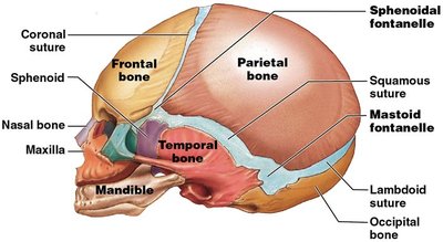
Skull
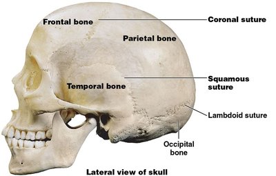
The skull is composed of two sets of bones: the cranium and the facial bones. The cranium encloses and protects the brain, while the facial bones form the structure of the face, hold the eyes, and provide attachment for facial muscles. The bones of the skull are joined by sutures, which are immovable joints, except for the mandible, which is attached by a freely movable joint.
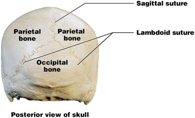
Cranial Bones (8): Frontal, occipital, ethmoid, sphenoid, two parietal, two temporal
Facial Bones (14): Maxillae (2), palatine (2), lacrimal (2), zygomatic (2), nasal (2), vomer, inferior nasal conchae (2), mandible
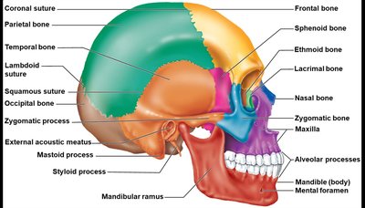
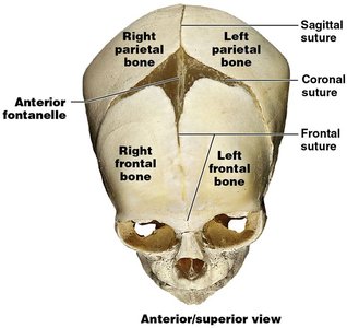
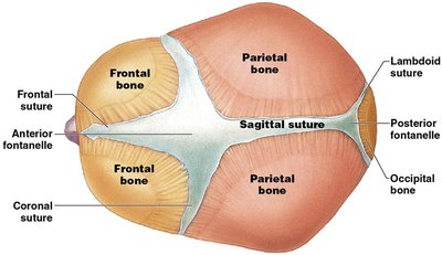
Sutures of the Skull
Sutures are immovable joints that connect the bones of the skull. They are held together by dense fibrous connective tissue. Major sutures include:
Coronal suture: Between frontal and parietal bones
Squamous suture: Between temporal and parietal bones
Lambdoid suture: Between occipital and parietal bones
Sagittal suture: Between the two parietal bones
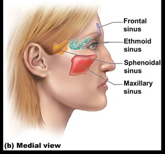
Paranasal Sinuses
Paranasal sinuses are hollow portions of certain skull bones surrounding the nasal cavity. Their functions include lightening the skull and amplifying sounds produced during speech.
Hyoid Bone
The hyoid bone is unique because it does not articulate with any other bone. It serves as a movable base for the tongue and aids in swallowing and speech.
Fontanelles
Fontanelles are soft spots on an infant's skull where the bones have not yet fused. They allow for cranial growth and ease passage through the birth canal. The anterior fontanelle is the largest and persists until about age 2. Other fontanelles (occipital, sphenoidal, mastoid) close within a few months after birth.
Vertebral Column
The vertebral column (spine) provides axial support, extending from the skull to the pelvis. It consists of 26 vertebrae separated by intervertebral discs:
7 cervical vertebrae (neck)
12 thoracic vertebrae (chest)
5 lumbar vertebrae (lower back)
Sacrum (fusion of 5 vertebrae)
Coccyx (fusion of 3–5 vertebrae)
Spinal curvatures are classified as:
Primary curvatures: Thoracic and sacral, present from birth (C-shaped in newborns)
Secondary curvatures: Cervical and lumbar, develop after birth (S-shaped in adults)
Features of Vertebrae
Each vertebra has several key features:
Body (centrum): Main weight-bearing region
Vertebral arch: Encloses the vertebral foramen
Pedicle and lamina: Parts of the vertebral arch
Processes: Transverse, spinous, superior and inferior articular processes
Thoracic Cage (Bony Thorax)
Structure and Function
The thoracic cage provides bony support to the walls of the thoracic cavity, protecting the heart, lungs, and thymus. It also serves as an attachment point for muscles involved in breathing, maintaining posture, and moving the upper limbs.
Sternum: Manubrium, body, xiphoid process
Ribs: 12 pairs (true ribs 1–7, false ribs 8–12, floating ribs 11–12)
Thoracic vertebrae
Appendicular Skeleton
Overview
The appendicular skeleton consists of 126 bones, including the limbs and the pectoral and pelvic girdles. It is responsible for facilitating movement and supporting the weight of the body during locomotion.
Pectoral (Shoulder) Girdle
The pectoral girdle attaches the upper limbs to the axial skeleton and consists of two clavicles and two scapulae. It is light and allows for a wide range of motion in the upper limbs.
Upper Limbs
Humerus: The single bone of the arm, articulates proximally with the scapula and distally with the radius and ulna.
Forearm: Composed of the ulna (medial) and radius (lateral).
Hand: Carpals (wrist, 8 bones), metacarpals (palm, 5 bones), phalanges (fingers, 14 bones per hand).
Pelvic Girdle
The pelvic girdle is formed by two hip bones (coxal bones), each resulting from the fusion of the ilium, ischium, and pubis. The pelvis supports the weight of the upper body and protects organs such as the reproductive organs, urinary bladder, and part of the large intestine.
Lower Limbs
Femur: Thigh bone, the heaviest and strongest bone in the body.
Patella: Sesamoid bone in the quadriceps tendon.
Leg: Tibia (medial, weight-bearing) and fibula (lateral, non-weight-bearing).
Foot: Tarsals (7 bones, including calcaneus and talus), metatarsals (5 bones), phalanges (14 bones).
Comparison of Male and Female Pelvis
The female pelvis is adapted for childbirth and differs from the male pelvis in several ways:
Larger and more circular pelvic inlet
Shallower and lighter bones
Ilia flare more laterally
Shorter, less curved sacrum
Wider pubic arch (≥ 100 degrees)
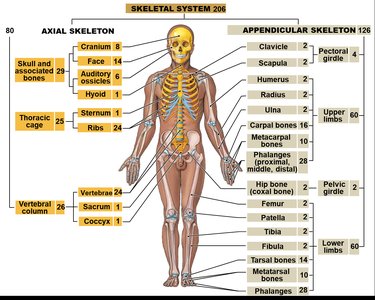
Summary Table: Major Bones of the Human Skeleton
Division | Bone Group | Number of Bones |
|---|---|---|
Axial Skeleton | Skull (Cranium + Face) | 22 |
Axial Skeleton | Auditory Ossicles | 6 |
Axial Skeleton | Hyoid | 1 |
Axial Skeleton | Vertebral Column | 26 |
Axial Skeleton | Thoracic Cage (Sternum + Ribs) | 25 |
Appendicular Skeleton | Pectoral Girdle (Clavicle + Scapula) | 4 |
Appendicular Skeleton | Upper Limbs | 60 |
Appendicular Skeleton | Pelvic Girdle | 2 |
Appendicular Skeleton | Lower Limbs | 60 |
Identify and label the bones of the axial and appendicular skeleton.
Identify cranial sutures and fontanelles, and explain the function of fontanelles.
Identify the components of the thoracic cage (sternum parts, types of ribs).
Explain the development and function of spinal curvatures.
Identify structural and functional differences between the male and female pelvis.
