 Back
BackThe Skeletal System: Structure, Classification, and Bone Markings
Study Guide - Smart Notes
 Tailored notes based on your materials, expanded with key definitions, examples, and context.
The Skeletal System
Functions of Bones
The skeletal system is essential for providing structural support, protecting internal organs, enabling movement, storing minerals and fats, and facilitating blood cell formation.
Support: Bones form the framework that supports the body.
Protection: Bones such as the skull and rib cage protect vital organs (e.g., brain, heart, lungs).
Movement: Skeletal muscles attach to bones, allowing movement at joints.
Storage: Bones store minerals (calcium, phosphorus) and fats (in marrow cavities).
Hematopoiesis: Blood cell formation occurs in the bone marrow.
Classification of Bones
Bones are classified by their shape and the type of tissue they contain. The adult skeleton consists of 206 bones, which are categorized as follows:
Compact bone: Dense, smooth, and homogeneous tissue.
Spongy bone: Composed of small needlelike pieces and many open spaces.
Bones are also classified by shape:
Long bones: Longer than wide, mostly compact bone (e.g., femur, humerus).
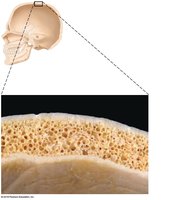
Flat bones: Thin, flattened, usually curved, with spongy bone sandwiched between compact bone.
Short bones: Cube-shaped, mostly spongy bone (e.g., wrist, ankle).
Irregular bones: Complex shapes (e.g., vertebrae).
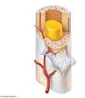
Structure of Long Bones
Long bones have a shaft (diaphysis) and two ends (epiphyses). The diaphysis is composed of compact bone, while the epiphyses contain spongy bone. The medullary cavity within the diaphysis contains yellow bone marrow in adults.
Periosteum: A fibrous membrane covering the bone.
Endosteum: Lines the medullary cavity.
Articular cartilage: Covers the epiphyses, reducing friction at joints.
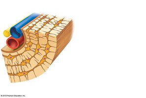
Microscopic Anatomy of Bone
Bone tissue is organized into structural units called osteons (Haversian systems). Compact bone contains concentric rings (lamellae) of matrix surrounding a central canal. Spongy bone is made of trabeculae and open spaces filled with marrow, blood vessels, and nerves.
Osteocytes: Mature bone cells located in lacunae.
Lacunae: Small cavities housing osteocytes.
Lamellae: Concentric circles of matrix.
Canaliculi: Tiny canals connecting lacunae.
Bone Markings
Projections and Processes
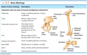
Bone markings are features that serve as sites for muscle and ligament attachment or as passageways for blood vessels and nerves. Projections and processes include:
Name | Description | Illustration |
|---|---|---|
Tuberosity | Large, rounded projection; may be rough | Femur |
Crest | Narrow ridge of bone; usually prominent | Iliac crest |
Trochanter | Very large, blunt, irregularly shaped process (only on femur) | Femur |
Line | Narrow ridge of bone; less prominent than crest | Femur |
Tubercle | Small, rounded projection or process | Humerus |
Epicondyle | Raised area on or above a condyle | Humerus |
Spine | Sharp, slender, often pointed projection | Vertebra |
Process | Any bony prominence | Vertebra |
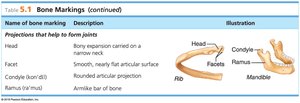
Projections That Help Form Joints
Name | Description | Illustration |
|---|---|---|
Head | Bony expansion carried on a narrow neck | Femur, humerus |
Facet | Smooth, nearly flat articular surface | Vertebra |
Condyle | Rounded articular projection | Mandible |
Ramus | Arm-like bar of bone | Mandible |
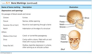
Depressions and Openings
Name | Description | Illustration |
|---|---|---|
Groove | Furrow | Skull |
Fissure | Narrow, slitlike opening | Skull |
Foramen | Round or oval opening through a bone | Skull |
Notch | Indentation at the edge of a structure | Skull |
Meatus | Canal or tunnel-like passageway | Skull |
Sinus | Cavity within a bone, filled with air and lined with mucous membrane | Skull |
Fossa | Shallow, basinlike depression in a bone, often serving as an articular surface | Skull |
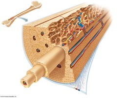
Microscopic Structure of Compact Bone
Osteon (Haversian System)
The osteon is the fundamental unit of compact bone, consisting of concentric lamellae surrounding a central canal. Blood vessels and nerves run through the central canal, supplying the bone tissue.
Perforating (Volkmann's) canals: Run perpendicular to the central canal, connecting blood and nerve supply.
Periosteum: The outer fibrous layer covering the bone.
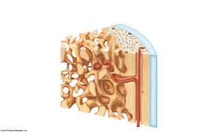
Microscopic Structure of Spongy Bone
Trabeculae and Open Spaces
Spongy bone is composed of trabeculae, which are small, needlelike pieces of bone. The open spaces between trabeculae are filled with bone marrow, blood vessels, and nerves, contributing to the lightweight nature of spongy bone.
Trabeculae: Provide structural support and house marrow.
Open spaces: Allow for the passage of blood vessels and nerves.
Summary Table: Bone Markings
Purpose of Table
This table summarizes the main types of bone markings, their descriptions, and their anatomical illustrations. Bone markings are essential for understanding muscle attachment, joint formation, and passageways for nerves and blood vessels.
Type | Description | Example |
|---|---|---|
Projection | Site of muscle/ligament attachment | Tuberosity, crest, trochanter |
Joint formation | Articular surface | Head, facet, condyle |
Depression/opening | Passage for vessels/nerves | Foramen, meatus, sinus |
