 Back
BackThe Skeletal System: Structure, Divisions, and Key Features
Study Guide - Smart Notes
 Tailored notes based on your materials, expanded with key definitions, examples, and context.
The Skeletal System
Overview of the Skeletal System
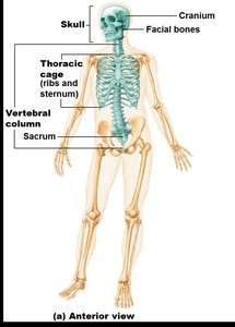
The skeletal system provides the structural framework for the human body, supports movement, protects internal organs, stores minerals, and produces blood cells. It is divided into two major divisions: the axial skeleton and the appendicular skeleton.
Axial Skeleton: Forms the longitudinal axis of the body and includes the skull, vertebral column, and bony thorax.
Appendicular Skeleton: Composed of the bones of the limbs and girdles that attach them to the axial skeleton.
Axial Skeleton
The Skull
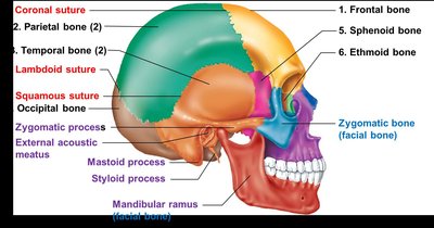
The skull is composed of two sets of bones: the cranium and the facial bones. The cranium protects the brain, while the facial bones form the structure of the face and provide cavities for the sense organs.
Cranium: Consists of 8 bones: frontal, parietal (2), occipital, temporal (2), sphenoid, and ethmoid.
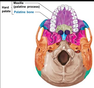
Facial Bones: Consists of 14 bones, including the mandible, maxillae, zygomatics, nasals, lacrimals, palatines, inferior nasal conchae, and vomer.
Sutures: Immovable joints that connect the bones of the skull (e.g., coronal, sagittal, lambdoid, squamous).
Mandible: The only freely movable bone of the skull.
Key Features of Cranial Bones
Zygomatic Process: Forms the cheekbone with the zygomatic bone.
External Acoustic Meatus: Canal for the external and middle ear.
Mastoid and Styloid Processes: Attachment sites for neck muscles.
Mandibular Ramus: Site for muscle attachment, blood vessels, and nerves.
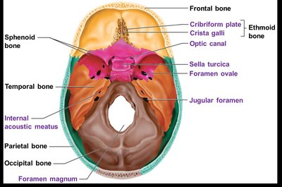
Internal Features of the Skull
Cribriform Plate (Ethmoid): Roof of the nasal cavity, passage for olfactory nerves.
Crista Galli (Ethmoid): Attachment for brain membranes.
Optic Canal (Sphenoid): Passage for the optic nerve.
Sella Turcica (Sphenoid): Houses the pituitary gland.
Foramen Magnum (Occipital): Exit for the spinal cord.
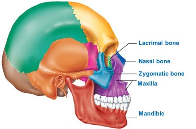
Facial Bones
The facial bones form the structure of the face, house the teeth, and provide attachment points for facial muscles.
Orbit: The eye socket is formed by seven bones: frontal, sphenoid, zygomatic, maxilla, lacrimal, ethmoid, and palatine.
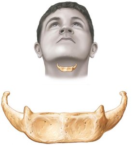
Hyoid Bone
The hyoid bone is unique because it does not articulate with any other bone. It serves as a movable base for the tongue and aids in swallowing and speech.
Paranasal Sinuses
Paranasal sinuses are air-filled spaces within certain skull bones that lighten the skull and amplify sounds produced during speech.
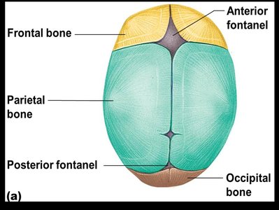
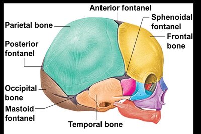
The Fetal Skull and Fontanels
The fetal skull is proportionally larger than the adult skull and contains fontanels—fibrous membranes between cranial bones that allow for compression during birth and brain growth during infancy. These fontanels ossify within 24 months after birth.
Fontanels: Anterior, posterior, sphenoidal, and mastoid.
Developmental Aspects: Cleft Palate
Cleft Palate: A congenital abnormality where the right and left halves of the palate do not fuse, resulting in a gap in the roof of the mouth. This can cause feeding and speech difficulties but can be repaired surgically.
Vertebral Column (Spine)
The vertebral column provides axial support, protects the spinal cord, and allows flexible movement. It consists of 26 vertebrae separated by intervertebral discs.
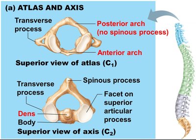
Cervical Vertebrae: 7 in the neck
Thoracic Vertebrae: 12 in the chest region
Lumbar Vertebrae: 5 in the lower back
Sacrum: Fusion of 5 vertebrae
Coccyx: Fusion of 3–5 vertebrae (tailbone)
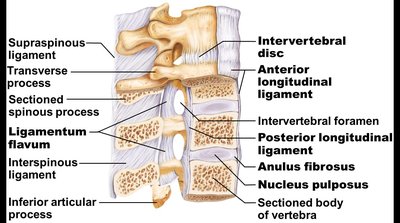
Ligaments of the Vertebral Column
Anterior and Posterior Longitudinal Ligaments: Hold the vertebral column in place from neck to sacrum.
Ligamentum Flavum: Connects adjacent vertebrae.
Short Ligaments: Connect each vertebra to those above and below.
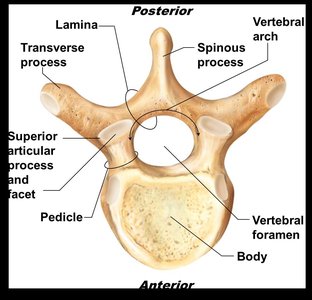
Structure of a Typical Vertebra
Body (Centrum): Weight-bearing region.
Vertebral Arch: Formed by pedicles and laminae, enclosing the vertebral foramen.
Vertebral Foramen: Passage for the spinal cord.
Processes: Transverse, spinous, superior and inferior articular processes for muscle attachment and articulation.
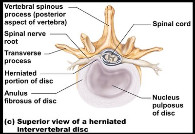
Intervertebral Discs
Nucleus Pulposus: Inner gelatinous core providing elasticity and compressibility.
Anulus Fibrosus: Outer collar of collagen and fibrocartilage.
Regional Characteristics of Vertebrae
Vertebrae differ in structure depending on their region (cervical, thoracic, lumbar) to accommodate different functions and ranges of motion.
Characteristic | Cervical (C3–7) | Thoracic | Lumbar |
|---|---|---|---|
Body | Small, wide side to side | Larger, heart-shaped | Massive, kidney-shaped |
Spinous process | Short, bifid | Long, projects inferiorly | Short, blunt, rectangular |
Vertebral foramen | Triangular | Circular | Triangular |
Transverse processes | Contain foramina | Bear facets for ribs | Thin and tapered |
Movements allowed | Flexion, extension, lateral flexion, rotation | Rotation, limited flexion/extension | Flexion, extension, some lateral flexion |
Curvatures of the Vertebral Column
Primary Curvatures: Thoracic and sacral, present from birth (C-shaped in newborns).
Secondary Curvatures: Cervical and lumbar, develop after birth (S-shaped in adults).
Abnormal Curvatures: Scoliosis (lateral), kyphosis (excessive thoracic), lordosis (excessive lumbar).
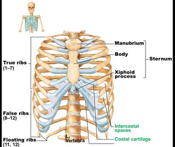
The Bony Thorax (Thoracic Cage)
The thoracic cage protects the heart and lungs and supports the shoulder girdles and upper limbs. It consists of the sternum, ribs, and thoracic vertebrae.
Sternum: Manubrium, body, xiphoid process.
Ribs: 12 pairs—true ribs (1–7), false ribs (8–12), floating ribs (11–12).
Thoracic Vertebrae: Posterior attachment for ribs.
Appendicular Skeleton
Overview
The appendicular skeleton consists of 126 bones, including the limbs and the pectoral and pelvic girdles, which attach the limbs to the axial skeleton.

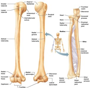
Upper Limb
Arm: Humerus
Forearm: Radius and ulna
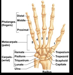
Hand: 8 carpals (wrist), 5 metacarpals (palm), 14 phalanges (fingers)
Lower Limb
Thigh: Femur
Leg: Tibia and fibula
Patella: Kneecap
Foot: 7 tarsals, 5 metatarsals, 14 phalanges
Pectoral (Shoulder) Girdle
Composed of: Clavicle and scapula
Function: Attaches the upper limb to the axial skeleton, allows a wide range of motion
Shoulder Joint: Formed by the head of the humerus sitting in the glenoid cavity of the scapula
Pelvic Girdle
Composed of: Two coxal bones (ilium, ischium, pubis), joined anteriorly at the pubic symphysis and posteriorly by the sacroiliac joint
Function: Supports the weight of the upper body, protects pelvic organs, forms the birth canal in females
Acetabulum: Socket for the head of the femur
Iliac Crest: Highest point of the pelvis (hips)
Gender Differences of the Pelvis
Female Pelvis: Larger, more circular inlet; shallower; lighter and thinner bones; iliac crests flare more; shorter, less curved sacrum; wider pubic arch.
Male Pelvis: Narrower, deeper, heavier bones; more acute pubic arch.
Summary of Learning Objectives
Identify the two major divisions of the skeletal system.
Know in detail all bones, bone features, unique characteristics, homeostatic imbalances, and diseases associated with the axial skeleton.
Know in detail all bones, bone features, unique characteristics, homeostatic imbalances, and diseases associated with the appendicular skeleton.
