 Back
BackThe Skeletal System: Structure, Divisions, and Key Features
Study Guide - Smart Notes
 Tailored notes based on your materials, expanded with key definitions, examples, and context.
The Skeletal System: General Organization
Overview of the Human Skeleton
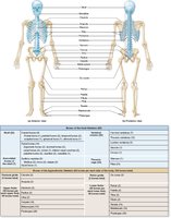
The human skeletal system provides structural support, protection for internal organs, and facilitates movement. It consists of 206 named bones, which are grouped into two principal divisions: the axial skeleton and the appendicular skeleton.
Axial skeleton: Composed of 80 bones, including the skull, vertebral column, and thoracic cage (ribs and sternum).
Appendicular skeleton: Composed of 126 bones, including the bones of the upper and lower limbs and the girdles (pectoral and pelvic) that attach them to the axial skeleton.
Divisions of the Skeletal System
Axial Skeleton
The axial skeleton forms the central axis of the body and is responsible for protecting the brain, spinal cord, and thoracic organs.
Skull: Protects the brain and forms the structure of the face.
Vertebral column: Supports the body and protects the spinal cord.
Thoracic cage: Includes the ribs and sternum, protecting the heart and lungs.
Hyoid bone: Supports the tongue and is not directly attached to other bones.
Appendicular Skeleton
The appendicular skeleton facilitates movement and interaction with the environment.
Pectoral girdle: Clavicle and scapula, attaching the upper limbs to the trunk.
Upper limbs: Humerus, radius, ulna, carpals, metacarpals, and phalanges.
Pelvic girdle: Hip bones (ossa coxae), attaching the lower limbs to the trunk.
Lower limbs: Femur, patella, tibia, fibula, tarsals, metatarsals, and phalanges.
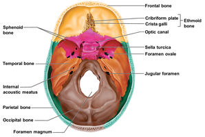
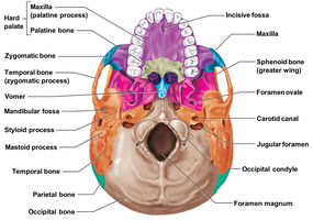
Skull: Cranial and Facial Bones
Major Bones of the Skull
The skull is composed of 22 bones, divided into cranial and facial bones. Cranial bones protect the brain, while facial bones form the structure of the face and house the special sense organs.
Cranial bones (8): Frontal, parietal (2), temporal (2), occipital, sphenoid, ethmoid.
Facial bones (14): Zygomatic (2), lacrimal (2), nasal (2), vomer, inferior nasal conchae (2), palatine (2), maxilla (2), mandible.
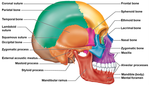
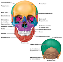
Key Features and Sutures of the Skull
Sutures are immovable joints that connect the bones of the skull. Major sutures include:
Coronal suture: Between frontal and parietal bones.
Sagittal suture: Between right and left parietal bones.
Squamous suture: Between parietal and temporal bones.
Lambdoid suture: Between parietal and occipital bones.
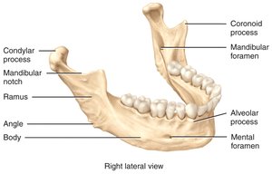
The Mandible
The mandible, or lower jawbone, is the largest and strongest facial bone. It holds the lower teeth and forms the only movable joint in the skull.
Key features: Body, ramus, angle, coronoid process, condylar process, mental foramen, mandibular foramen, alveolar process.
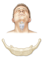
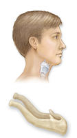
Hyoid Bone
Structure and Function
The hyoid bone is a slender, U-shaped bone located in the anterior neck. It does not articulate with any other bone and serves as an attachment site for tongue and neck muscles.
Parts: Body, greater cornu, lesser cornu.
Function: Supports the tongue and assists in swallowing and speech.
Vertebral Column
General Structure and Function
The vertebral column, or spine, is composed of 24 individual vertebrae and two fused bones (sacrum and coccyx). It provides vertical support, protects the spinal cord, and allows flexible movement.
Functions: Supports the head, maintains upright posture, protects the spinal cord, and serves as an attachment for ribs and muscles.
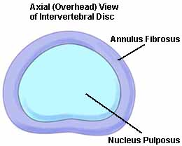
Intervertebral Discs
Intervertebral discs are pads of fibrocartilage located between vertebral bodies. They act as shock absorbers and allow flexibility in the spine.
Structure: Annulus fibrosus (outer ring) and nucleus pulposus (inner core).
Divisions of the Vertebral Column
The vertebral column is divided into five regions, each with distinct characteristics and functions.
Cervical (C1–C7): Neck region, supports the head.
Thoracic (T1–T12): Upper back, articulates with ribs.
Lumbar (L1–L5): Lower back, bears most body weight.
Sacral (S1–S5): Fused to form the sacrum.
Coccygeal (Co1–Co4): Fused to form the coccyx (tailbone).
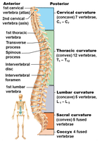
Vertebral Curvatures
The vertebral column has four main curvatures that help distribute weight and maintain balance.
Primary curves: Thoracic and sacral (present at birth, C-shaped).
Secondary curves: Cervical and lumbar (develop after birth, S-shaped).
Types of Vertebrae
Cervical vertebrae (C1–C7): Smallest, with transverse foramina. Atlas (C1) allows nodding, axis (C2) allows rotation.
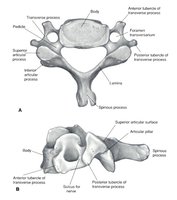
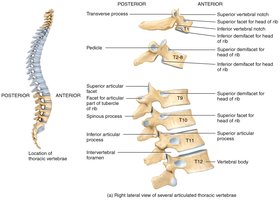
Thoracic vertebrae (T1–T12): Articulate with ribs, have long spinous processes.
Lumbar vertebrae (L1–L5): Largest, support most weight, have thick bodies.
Summary Table: Axial vs. Appendicular Skeleton
Division | Major Components | Number of Bones | Key Functions |
|---|---|---|---|
Axial Skeleton | Skull, vertebral column, thoracic cage, hyoid bone | 80 | Protection, support, central axis |
Appendicular Skeleton | Pectoral girdle, upper limbs, pelvic girdle, lower limbs | 126 | Movement, interaction with environment |
Key Terms and Definitions
Axial skeleton: The central part of the skeleton, including the skull, vertebral column, and thoracic cage.
Appendicular skeleton: The bones of the limbs and girdles that attach them to the axial skeleton.
Suture: An immovable joint between skull bones.
Intervertebral disc: A fibrocartilaginous pad between vertebrae, providing cushioning and flexibility.
Mandible: The lower jawbone, the only movable bone of the skull.
Hyoid bone: A U-shaped bone in the neck that does not articulate with other bones.
