 Back
BackThe Skeletal System: Structure, Function, and Anatomy
Study Guide - Smart Notes
 Tailored notes based on your materials, expanded with key definitions, examples, and context.
The Skeletal System
Overview and Components
The skeletal system is a complex framework of bones and connective tissues that provides structure, protection, and movement for the human body. It consists of bones, cartilages, joints, ligaments, and other connective tissues that stabilize or connect bones.
Bones: Rigid organs that form the skeleton.
Cartilages: Flexible connective tissues at joints and in some structures.
Joints: Articulations where two or more bones meet.
Ligaments: Connective tissues that connect bones to other bones.
Five Main Functions of the Skeletal System
Support: Provides structural support for the entire body and attachment points for soft tissues and organs.
Storage of Minerals and Lipids: Stores calcium and phosphorus ions in bone matrix; yellow bone marrow stores lipids for energy.
Blood Cell Production: Red bone marrow produces red blood cells, white blood cells, and platelets.
Protection: Surrounds and protects vital organs (e.g., skull protects the brain, ribs protect the heart and lungs).
Leverage: Bones act as levers to change the direction and magnitude of muscle forces, enabling movement.
Bone Tissue and Structure
Bone (Osseous) Tissue Characteristics
Bone tissue is a specialized connective tissue composed of cells and a mineralized matrix. The main cell type is the osteocyte.
Matrix: Contains calcium phosphate (Ca3(PO4)2) and collagen fibers.
Calcium salts provide hardness; collagen fibers provide flexibility and tensile strength.
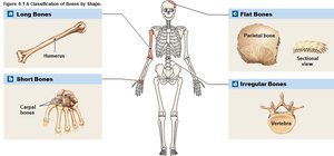
Classification of Bones by Shape
Bones are classified based on their shapes:
Long bones: Longer than they are wide (e.g., humerus).
Short bones: About as long as they are wide (e.g., carpal bones).
Flat bones: Thin and broad (e.g., parietal bone of the skull).
Irregular bones: Complex shapes (e.g., vertebrae).
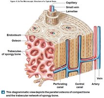
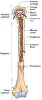
Features of a Long Bone
Diaphysis: Central shaft, surrounds the marrow (medullary) cavity.
Epiphyses: Expanded ends, covered with articular cartilage, articulate with adjacent bones.
Marrow cavity: Contains bone marrow (red or yellow).
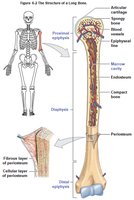
Types of Bone Tissue in a Long Bone
Compact bone (dense bone): Forms the wall of the diaphysis; solid and strong.
Spongy bone (trabecular bone): Network of bony rods separated by spaces; fills epiphyses and lines the marrow cavity.

Coverings of a Long Bone
Periosteum: Outer covering; has an outer fibrous layer and an inner cellular layer; isolates bone and provides attachment for tendons and ligaments.
Endosteum: Lines the marrow cavity and covers spongy bone; active in bone growth, repair, and remodeling.

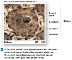
Microscopic Features of Bone
Osteocytes: Mature bone cells located in lacunae between lamellae (layers of matrix).
Canaliculi: Small channels connecting lacunae, allowing for nutrient and waste exchange.
Osteon (Haversian system): Basic functional unit of compact bone; concentric lamellae around a central canal containing blood vessels.

Spongy Bone Structure and Function
No osteons; lamellae form trabeculae (rods/plates).
Spaces between trabeculae contain red bone marrow.
Found in areas with multidirectional stress (e.g., epiphyses of long bones).
Lighter than compact bone, reducing skeletal weight.
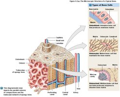
Bone Cells
Osteoblasts: Produce new bone matrix (ossification).
Osteocytes: Maintain bone matrix and recycle calcium salts.
Osteoclasts: Break down bone matrix (osteolysis/resorption).
Bone Formation and Growth
Ossification
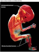
Ossification is the process of replacing other tissues with bone. It begins during embryonic development and continues into adolescence.
Intramembranous ossification: Bone develops from fibrous connective tissue (e.g., flat bones of the skull).
Endochondral ossification: Bone replaces a hyaline cartilage model (most bones form this way).
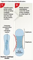
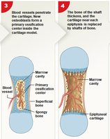
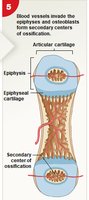
Endochondral Ossification Steps
Chondrocytes enlarge, matrix calcifies, and chondrocytes die.
Bone formation begins at the shaft surface as osteoblasts produce bone matrix.
Blood vessels invade cartilage; osteoblasts form spongy bone at the primary ossification center.
Bone develops toward the ends; osteoclasts create the marrow cavity.
Secondary ossification centers form in the epiphyses; articular cartilage remains at joint surfaces.
Epiphyseal Line and Growth in Length
Epiphyseal cartilage (growth plate) allows for bone lengthening during childhood and adolescence.
At puberty, increased sex hormones accelerate ossification, leading to epiphyseal closure and formation of the epiphyseal line (no further lengthening).
Appositional Growth (Bone Diameter)
Bones grow in diameter as osteoblasts add matrix to the outer surface and osteoclasts remove matrix from the inner surface, enlarging the marrow cavity.

Requirements for Bone Growth
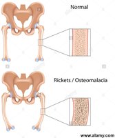
Vitamin D3: Needed for calcium absorption; deficiency causes osteomalacia (adults) or rickets (children).
Vitamins A and C: Support osteoblast function; vitamin C deficiency causes scurvy.
Minerals: Calcium and phosphate are essential for bone matrix.
Hormones: Growth hormone, thyroid hormones, sex hormones, and calcium-regulating hormones are all involved in bone growth and maintenance.
Bone Remodeling and Homeostasis
Bone Remodeling
Continuous process of bone matrix renewal and recycling by osteocytes, osteoblasts, and osteoclasts.
Allows bones to adapt to new stresses; exercise increases bone strength, while inactivity leads to bone loss.
Remodeling rate varies by age and bone type; spongy bone remodels more frequently than compact bone.
Calcium Homeostasis
99% of body calcium is stored in bones.
Calcium is essential for nerve and muscle function; blood calcium levels are tightly regulated.
Parathyroid hormone (PTH) and calcitriol increase blood calcium; calcitonin lowers blood calcium.
Bone Fractures and Repair
Types of Fractures
Closed (simple) fracture: Bone does not break the skin.
Open (compound) fracture: Bone pierces the skin; higher risk of infection.
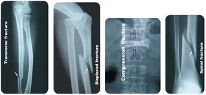
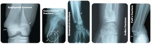
Other types: transverse, displaced, compression, spiral, epiphyseal, comminuted, greenstick, Colles, Pott's fracture.
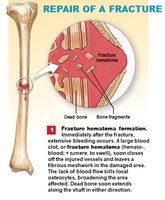
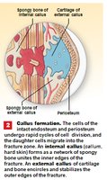
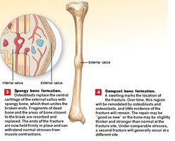
Fracture Repair Process
Formation of a fracture hematoma (blood clot).
Formation of an internal callus (spongy bone) and external callus (cartilage and bone).
Replacement of cartilage with spongy bone by osteoblasts.
Remodeling of bone; compact bone replaces spongy bone, leaving a thickened area at the fracture site.
Aging and Bone Health
Osteopenia and Osteoporosis
Osteopenia: Age-related reduction in bone mass; begins between ages 30–40.
Osteoporosis: Severe bone loss that impairs function and increases fracture risk; more common in women, especially after menopause due to decreased estrogen.
Bone Markings and Skeletal Divisions
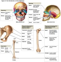
Bone Markings
Bone markings are surface features that serve as attachment sites for muscles, ligaments, and tendons, or as passages for nerves and blood vessels.
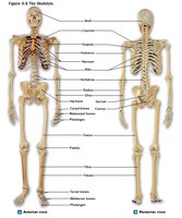
Skeletal Divisions
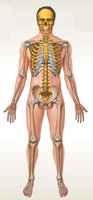
Axial skeleton: 80 bones including the skull, vertebral column, and thoracic cage.
Appendicular skeleton: 126 bones including the pectoral girdle, pelvic girdle, and limbs.
Summary Table: Types of Bone Cells
Cell Type | Function |
|---|---|
Osteoblast | Builds new bone matrix (ossification) |
Osteocyte | Maintains bone matrix, recycles calcium salts |
Osteoclast | Breaks down bone matrix (osteolysis) |
Additional info: This guide covers the essential structure and function of the skeletal system, bone tissue, bone growth and remodeling, fracture repair, and the organization of the human skeleton. For more detailed study, refer to textbook chapters on the skeletal system and related laboratory exercises.
