 Back
BackThe Skeletal System: Structure, Function, and Bone Anatomy
Study Guide - Smart Notes
 Tailored notes based on your materials, expanded with key definitions, examples, and context.
The Skeletal System: Structure, Function, and Bone Anatomy
Introduction to the Skeletal System
The skeletal system is a dynamic network of tissues that provides structural support, protection, and a framework for the body. It is essential for movement, storage of minerals, production of blood cells, and interaction with other organ systems. The shape and size of the skeleton largely determine the overall form of the human body.
Bone Terminology and Cell Types
Key Terms and Definitions
Osteocytes: Mature bone cells that maintain the bone matrix.
Osteoblasts: Bone-forming cells responsible for synthesizing and secreting the bone matrix.
Osteoclasts: Large cells that break down bone tissue, aiding in bone remodeling and calcium release.
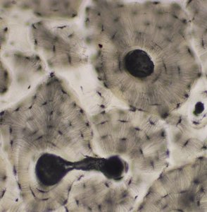
Lacunae: Small spaces within the bone matrix that house osteocytes.
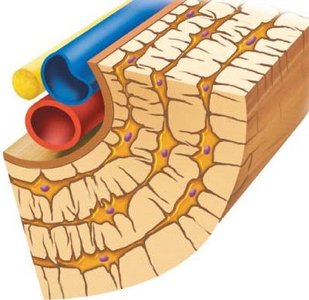
Lamellae: Layers of bone matrix; can be concentric (around a central canal), interstitial (between osteons), or circumferential (around the entire bone).
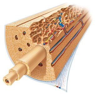
Haversian (central) canal: Central channel in an osteon containing blood vessels and nerves.
Volkmann’s (perforating) canal: Canals that run perpendicular to Haversian canals, connecting them and facilitating vascular and nerve supply.
Canaliculi: Tiny channels connecting lacunae, allowing for nutrient and waste exchange between osteocytes.
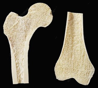
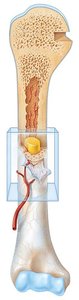
Diaphysis: The shaft of a long bone.
Epiphysis: The end part of a long bone, usually wider than the shaft.
Spongy (Cancellous) bone: Porous bone tissue found at the ends of long bones and inside flat bones.
Compact bone: Dense bone tissue forming the outer layer of bones.
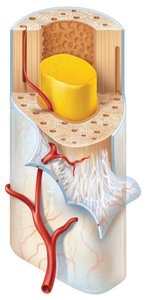
Medullary cavity: Central cavity within the diaphysis, containing bone marrow.
Red bone marrow: Hematopoietic tissue responsible for blood cell production.
Yellow bone marrow: Fatty tissue found in the medullary cavity of adults.
Endosteum: Thin membrane lining the medullary cavity.
Periosteum: Dense connective tissue covering the outer surface of bones.
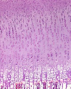
Epiphyseal plate: Growth plate made of cartilage, responsible for lengthwise bone growth in children and adolescents.
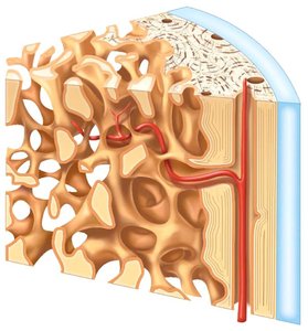
Bone Structure and Microscopic Features
Gross Anatomy of Bone
Bones are composed of both compact and spongy tissue, each serving distinct structural and functional roles. The diaphysis forms the shaft, while the epiphyses are the expanded ends. The medullary cavity within the diaphysis contains bone marrow.
Microscopic Anatomy of Compact Bone
Compact bone is organized into osteons (Haversian systems), which are cylindrical structures that provide strength. Each osteon consists of concentric lamellae surrounding a central canal. Osteocytes reside in lacunae and communicate via canaliculi.
Internal Features and Vascularization
The internal structure of bone includes a network of blood vessels, nerves, and marrow. The periosteum covers the outer surface, while the endosteum lines the inner surfaces. Spongy bone contains trabeculae, which are oriented along lines of stress to provide support.
Long Bone Structure
Long bones have a characteristic structure with a diaphysis, two epiphyses, and a medullary cavity. The medullary cavity contains yellow marrow in adults, and the bone is richly supplied with blood vessels and nerves.
Bone Matrix Composition
Organic vs. Inorganic Matrix
Organic matrix: Composed mainly of collagen fibers and ground substance, providing flexibility and tensile strength.
Inorganic matrix: Consists primarily of hydroxyapatite (calcium phosphate crystals), giving bone its hardness and resistance to compression.
Formula for hydroxyapatite:
Bone Formation and Growth
Types of Bone Formation
Intramembranous ossification: Bone develops directly from mesenchymal tissue (e.g., flat bones of the skull).
Endochondral ossification: Bone forms by replacing hyaline cartilage (e.g., long bones).
Endochondral Ossification and Growth Plates
Postnatal bone growth in length occurs at the epiphyseal plate through interstitial growth. This process involves several zones, each with distinct cellular activities, from cartilage proliferation to ossification.
Bone Markings and Terminology
General Bone Markings
Process: Any bony projection
Condyle: Rounded articulating process
Epicondyle: Projection above a condyle
Tuberosity: Large, rounded or irregular process
Tubercle: Small, rounded process
Trochanter: Very large, blunt process (femur only)
Spine: Sharp, slender process
Hamulus: Hook-shaped process
Line: Slight ridge of bone
Crest: Prominent ridge of bone
Facet: Smooth, flattened articulating surface
Fossa: Bony depression
Foramen: Hole in a bone for passage of nerves/blood vessels
Meatus/Canal: Tunnel-like passage
Sinus: Cavity within a bone
Sulcus/Groove: Furrow on a bone's surface
Fissure: Slit-like opening
Fovea: Shallow depression
Axial and Appendicular Skeleton Overview
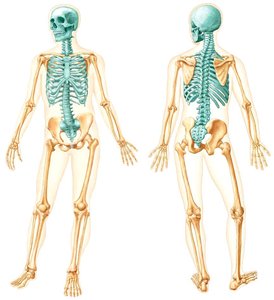
Axial Skeleton
The axial skeleton includes the skull, vertebral column, and thoracic cage. It provides central support and protects vital organs.
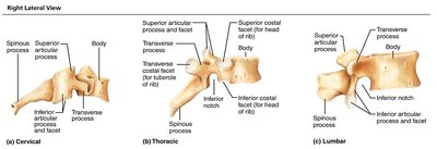
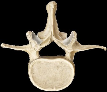
Vertebral Column
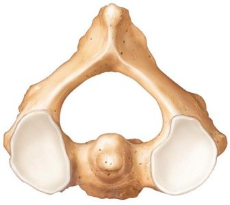
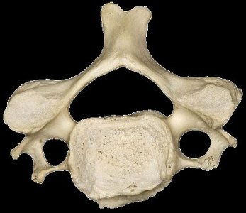
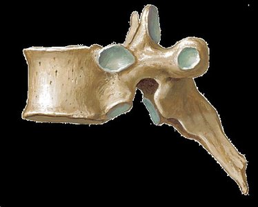
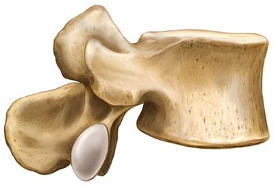
The vertebral column consists of cervical, thoracic, lumbar, sacral, and coccygeal regions. Each vertebra has characteristic features such as the body, vertebral foramen, spinous process, and transverse process.
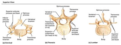
Specialized Vertebrae
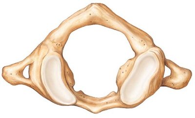
Atlas (C1): Supports the skull, allows nodding motion.
Axis (C2): Has the odontoid process (dens) for rotation of the head.
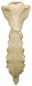
Sacrum and Coccyx
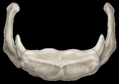
The sacrum is a triangular bone formed by the fusion of five sacral vertebrae, while the coccyx is formed by the fusion of four small vertebrae.
Thoracic Cage
The thoracic cage consists of the sternum, ribs, and thoracic vertebrae. It protects the heart and lungs and supports the shoulder girdles.
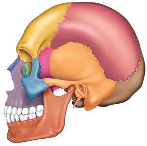
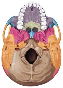
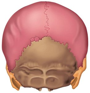
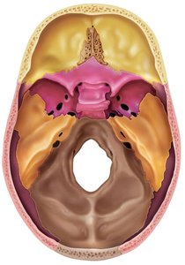
Skull Anatomy
The skull is composed of cranial and facial bones, with numerous markings and sutures. It protects the brain and forms the structure of the face.

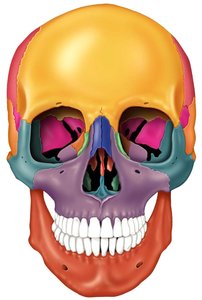
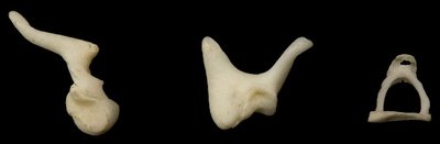
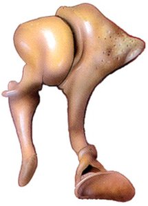
Auditory Ossicles and Hyoid Bone
The auditory ossicles (malleus, incus, stapes) are small bones in the middle ear, while the hyoid bone supports the tongue and is not directly attached to other bones.
Appendicular Skeleton Overview
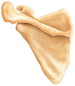
Pectoral Girdle and Upper Limb
The pectoral girdle consists of the clavicle and scapula, providing attachment for the upper limb. The upper limb includes the humerus, radius, ulna, carpals, metacarpals, and phalanges.
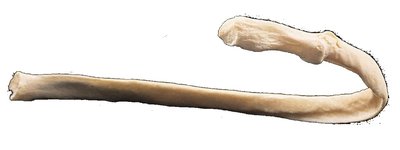
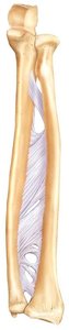
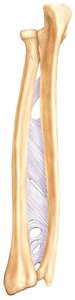
Radius and Ulna
The radius and ulna are the two bones of the forearm, connected by an interosseous membrane. They articulate with the humerus proximally and the carpal bones distally.
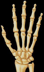
Wrist and Hand
The wrist contains eight carpal bones, while the hand includes five metacarpals and fourteen phalanges.
Pelvic Girdle and Lower Limb
The pelvic girdle is formed by the os coxae (ilium, ischium, pubis). The lower limb includes the femur, tibia, fibula, patella, tarsals, metatarsals, and phalanges.
Summary Table: Differences Between Cartilage and Bone
Feature | Cartilage | Bone |
|---|---|---|
Nerves | Absent | Present |
Blood vessels | Absent | Present |
Lymph channels | Absent | Present |
Matrix type | Gel-like, rich in proteoglycans | Hard, rich in hydroxyapatite |
Example: The femur is a long bone with a diaphysis, two epiphyses, a medullary cavity containing yellow marrow, and both compact and spongy bone tissue. It articulates with the pelvis at the hip and the tibia at the knee.
Additional info: Bone is a highly vascularized tissue, which allows it to heal more rapidly than cartilage. The presence of nerves and blood vessels in bone is essential for nutrient delivery, waste removal, and sensory function.
