 Back
BackThe Skeletal System: Structure, Function, and Clinical Relevance
Study Guide - Smart Notes
 Tailored notes based on your materials, expanded with key definitions, examples, and context.
Skeletal System Overview
Functions of the Skeletal System
The skeletal system provides the essential framework for the human body, supporting and protecting organs, enabling movement, and serving as a reservoir for minerals and blood cell production.
Support: Forms the internal framework that supports the body.
Protection: Shields internal organs and aids in immune defense by producing white blood cells.
Movement: Facilitates movement by serving as levers for muscle action.
Mineral Storage: Stores calcium and phosphorus, vital for physiological processes.
Hematopoiesis: Produces blood cells in red bone marrow.
Classification and Structure of Bones
Types of Bones
Bones are classified by shape, which relates to their function and location in the body.
Long Bones: Longer than they are wide (e.g., humerus).
Short Bones: Cube-shaped (e.g., carpals of the wrist).
Flat Bones: Thin and often curved (e.g., frontal bone of the skull).
Irregular Bones: Complex shapes (e.g., vertebrae).
Sesamoid Bones: Develop within tendons (e.g., patella).
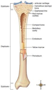
Structure of Long Bones
Long bones have a specialized structure that supports their function in movement and weight-bearing.
Diaphysis: The shaft, composed of compact bone.
Medullary Cavity: Central cavity containing yellow marrow (fat storage).
Epiphyses: Ends of the bone, made of spongy bone containing red marrow.
Articular Cartilage: Covers joint surfaces, reducing friction and absorbing shock.
Periosteum: Tough membrane covering the bone except at joints; contains nerves and blood vessels.
Endosteum: Thin membrane lining the medullary cavity.
Structure of Flat Bones
Flat bones consist of a layer of spongy bone (diploë) sandwiched between two layers of compact bone, providing protection and broad surfaces for muscle attachment.

Microscopic Structure of Bone
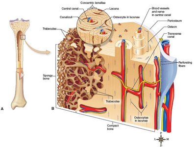
Bone Tissue Types
Spongy (Cancellous) Bone: Composed of trabeculae with spaces containing marrow; found in epiphyses.
Compact Bone: Dense and organized into osteons (Haversian systems) for strength.
Osteon (Haversian System)
The osteon is the fundamental unit of compact bone, consisting of concentric lamellae, lacunae (housing osteocytes), canaliculi (nutrient channels), and a central canal with blood vessels.
Cartilage Tissue Structure
Chondrocytes: Cartilage cells located in lacunae.
Matrix: Flexible, gel-like, and avascular (lacks blood vessels).
Bone Development and Growth
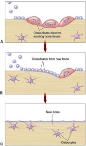
Bone Cells
Osteoblasts: Bone-forming cells.
Osteoclasts: Bone-resorbing cells.
Osteocytes: Mature bone cells maintaining bone tissue.
Ossification Processes
Endochondral Ossification: Most bones form from cartilage models; ossification centers appear in the diaphysis and epiphyses.
Intramembranous Ossification: Flat bones form within connective tissue membranes.
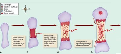
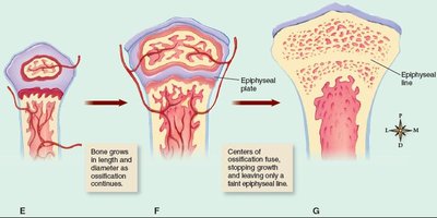
Bone Development in Infancy
At birth, the skeleton consists largely of cartilage and fibrous structures, which are gradually replaced by bone through ossification.
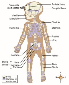
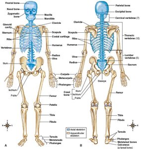
Divisions of the Skeleton
Axial and Appendicular Skeleton
Axial Skeleton (80 bones): Skull, vertebral column, thorax.
Appendicular Skeleton (126 bones): Upper and lower limbs, pectoral and pelvic girdles.
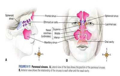
Skull
8 cranial bones, 14 facial bones, 6 middle ear bones.
Sinuses lighten the skull and enhance resonance of voice.
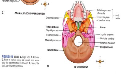
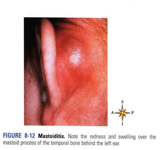
Mastoiditis
Inflammation of the mastoid air cells in the temporal bone can lead to serious complications if untreated.
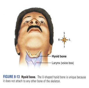
Hyoid Bone
The hyoid bone is unique as it does not articulate with any other bone and serves as an anchor for tongue muscles and supports the larynx.
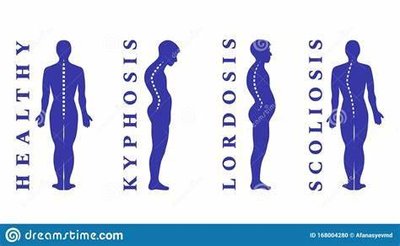
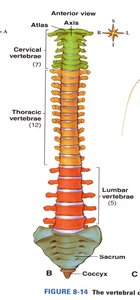
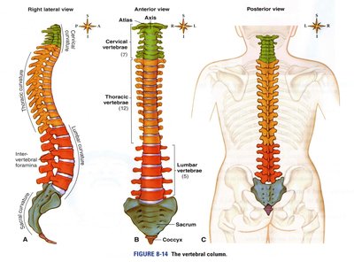
Vertebral Column
Composed of cervical (7), thoracic (12), lumbar (5), sacrum (1), and coccyx (1) bones.
Normal curves provide strength and flexibility; abnormal curves include lordosis, kyphosis, and scoliosis.
Name | Number | Description |
|---|---|---|
Cervical | 7 | Upper 7 vertebrae; first is atlas, second is axis |
Thoracic | 12 | Next 12 vertebrae; ribs attach here |
Lumbar | 5 | Lower back vertebrae |
Sacrum | 1 | Fused from 5 vertebrae in adults |
Coccyx | 1 | Fused from 3-5 vertebrae in adults |
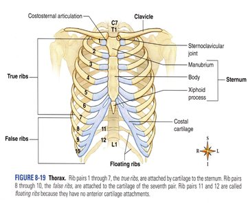
Thorax
Composed of 12 pairs of ribs, sternum, and thoracic vertebrae.
True ribs (1-7), false ribs (8-10), floating ribs (11-12).
Name | Number | Description |
|---|---|---|
True ribs | 14 | Upper seven pairs; attached to sternum by costal cartilages |
False ribs | 10 | Lower five pairs; lowest two are floating ribs |
Sternum | 1 | Breastbone; includes manubrium and xiphoid process |
Appendicular Skeleton
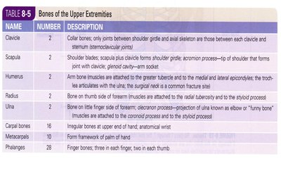
Upper Extremity: Clavicle, scapula, humerus, radius, ulna, carpals, metacarpals, phalanges.
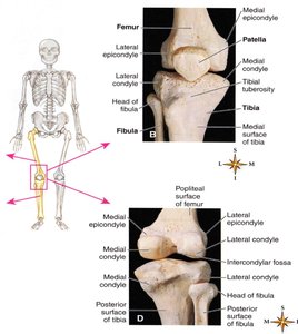
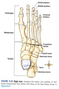
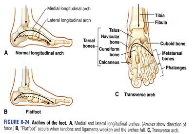
Lower Extremity: Coxal bone, femur, patella, tibia, fibula, tarsals, metatarsals, phalanges.
Name | Number | Description |
|---|---|---|
Clavicle | 2 | Collar bones; only joints between shoulder girdle and axial skeleton |
Scapula | 2 | Shoulder blades; forms shoulder girdle with clavicle |
Humerus | 2 | Upper arm bone |
Radius | 2 | Forearm bone on thumb side |
Ulna | 2 | Forearm bone on little finger side |
Carpals | 16 | Wrist bones |
Metacarpals | 10 | Palm bones |
Phalanges | 28 | Finger bones |
Name | Number | Description |
|---|---|---|
Coxal bone | 2 | Hip bones; each formed by fusion of ilium, ischium, and pubis |
Femur | 2 | Thigh bones |
Patella | 2 | Kneecap |
Tibia | 2 | Shinbone |
Fibula | 2 | Slender lateral bone of leg |
Tarsal bones | 14 | Ankle bones |
Metatarsals | 10 | Foot bones |
Phalanges | 28 | Toe bones |
Sexual and Age Differences in the Skeleton
Male Skeleton: Generally larger, pelvis deep and narrow, smaller pelvic inlet, narrower pubic angle.
Female Skeleton: Broader and shallower pelvis, wider pelvic inlet and pubic angle for childbirth.
Age: Skeleton matures around age 25; bone density decreases after age 50.
Joints (Articulations)
Classification by Movement
Synarthrosis: Immovable joints (e.g., skull sutures).
Amphiarthrosis: Slightly movable joints (e.g., symphysis pubis).
Diarthrosis: Freely movable joints (e.g., shoulder, knee).
Types of Diarthrotic Joints
Ball-and-Socket: Shoulder, hip.
Hinge: Elbow, knee.
Pivot: Atlas and axis of neck.
Saddle: Thumb joint.
Gliding: Intercarpal joints.
Condyloid: Wrist joint.
Structure of a Diarthrotic Joint
Joint Capsule and Ligaments: Hold bones together while permitting movement.
Articular Cartilage: Covers bone ends, absorbs shock.
Synovial Membrane: Secretes lubricating fluid.
Joint Cavity: Space between articulating bones.
Clinical Conditions of the Skeletal System
Bone and Cartilage Tumors
Osteosarcoma: Malignant bone tumor, often in distal femur, proximal tibia, or humerus.
Chondrosarcoma: Malignant cartilage tumor, often in humerus, femur, ribs, pelvis.
Metabolic Bone Diseases
Osteoporosis: Loss of bone matrix and trabeculae, leading to fractures; treated with drugs, exercise, calcium, and vitamin D.
Rickets: Loss of bone minerals in children, causing deformities; treated with vitamin D.
Osteomalacia: Loss of minerals in mature bones, increasing fracture risk; treated with vitamin D.
Paget Disease: Faulty bone remodeling, causing deformities and fractures; may be genetic or viral in origin.
Osteogenesis Imperfecta: Brittle bone disease due to lack of organic matrix; managed with splinting and medication.
Bone Infection
Osteomyelitis: Bacterial infection of bone, often requiring surgery and antibiotics.
Bone Fractures
Open (Compound) Fracture: Bone pierces the skin.
Closed (Simple) Fracture: Bone does not pierce the skin.
Other types: Complete, incomplete, linear, transverse, oblique.
Joint Conditions
Osteoarthritis (DJD): Degenerative, noninflammatory joint disease; most common cause for joint replacements.
Dislocation/Subluxation: Bones in a joint lose proper contact.
Sprain: Ligament injury.
Strain: Injury to muscle or tendon.
Arthritis: Inflammatory joint conditions, including rheumatoid arthritis (autoimmune), gouty arthritis (urate crystals), and infectious arthritis (bacterial).
Review Questions
What substance is stored in bones? Calcium
Red bone marrow fills in small spaces in the spongy bone composing the: Epiphyses
Osteocytes and chondrocytes lie within little spaces called: Lacunae
Bone-forming cells are called osteoblasts, whereas bone-resorbing cells are called osteoclasts.
Which of these bones would be found in the axial division of the skeleton? Ribs
The normal curves of the spine provide us with enough strength to support our weight and: Provide the balance necessary for us to stand upright
An example of an amphiarthrotic joint is: Symphysis pubis
A skeletal disorder that involves the demineralization of developing bones in infants and young children before skeletal maturity is: Rickets
