 Back
The Skeletal System: Structure, Function, and Disorders
Study Guide - Smart Notes
 Tailored notes based on your materials, expanded with key definitions, examples, and context.
Skeletal System Overview
General Functions of the Skeletal System
The skeletal system provides the essential framework for the human body, supporting and protecting organs, enabling movement, and serving as a reservoir for minerals and blood cell production.
Support: The skeleton forms the internal framework that supports the body and maintains its shape.
Protection: Bones protect vital organs (e.g., skull protects the brain, rib cage protects the heart and lungs).
Movement: Bones act as levers and points of attachment for muscles, facilitating movement.
Mineral Storage: Bones store calcium and phosphorus, releasing them as needed for physiological processes.
Hematopoiesis: Blood cell formation occurs in red bone marrow, a process known as hematopoiesis.
Classification and Structure of Bones
Types of Bones
Bones are classified by shape, which relates to their function and location in the body.
Long Bones: Longer than they are wide (e.g., humerus, femur).
Short Bones: Approximately equal in length and width (e.g., carpals, tarsals).
Flat Bones: Thin and often curved (e.g., frontal bone, sternum).
Irregular Bones: Complex shapes (e.g., vertebrae).
Sesamoid Bones: Small, round bones embedded in tendons (e.g., patella).
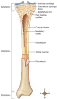
Structure of a Long Bone
Long bones have a specialized structure that supports their function in movement and weight-bearing.
Diaphysis: The shaft, composed of compact bone, provides strength.
Epiphyses: The ends of the bone, made of spongy bone containing red marrow.
Medullary Cavity: Central cavity containing yellow marrow (fat storage).
Articular Cartilage: Covers joint surfaces, reducing friction and absorbing shock.
Periosteum: Tough membrane covering the bone except at joints; contains nerves and blood vessels.
Endosteum: Thin membrane lining the medullary cavity.
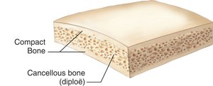
Structure of Flat Bones
Flat bones consist of a layer of spongy bone (diploë) sandwiched between two layers of compact bone, providing protection and a broad surface for muscle attachment.
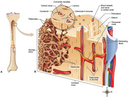
Microscopic Structure of Bone and Cartilage
Bone Tissue Types
Compact Bone: Dense and strong, composed of structural units called osteons (Haversian systems).
Spongy Bone: Contains trabeculae with spaces filled by red marrow; found mainly in epiphyses.
Osteon (Haversian System)
The osteon is the fundamental unit of compact bone, consisting of concentric lamellae, lacunae (housing osteocytes), canaliculi (tiny canals), and a central canal containing blood vessels.
Cartilage Tissue Structure
Chondrocytes: Cartilage cells located in lacunae.
Matrix: Flexible, gel-like, and avascular (lacks blood vessels), making cartilage slower to heal than bone.
Bone Formation, Growth, and Remodeling
Bone Cells and Remodeling
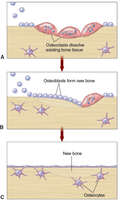
Bone is a dynamic tissue, constantly being formed and resorbed by specialized cells.
Osteoblasts: Cells that build new bone matrix.
Osteoclasts: Cells that break down bone matrix.
Osteocytes: Mature bone cells derived from osteoblasts, maintaining bone tissue.
Bone Formation Processes
Endochondral Ossification: Most bones form from cartilage models, which are gradually replaced by bone tissue.
Intramembranous Ossification: Some flat bones (e.g., skull) form directly from connective tissue membranes.
Endochondral Ossification Sequence
Bone development begins in the diaphysis (primary ossification center) and later in the epiphyses (secondary centers). The epiphyseal plate allows for longitudinal growth until it is replaced by bone (epiphyseal line) at maturity.
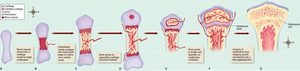
Bone Development in Newborns
At birth, much of the skeleton is still cartilage or fibrous tissue, which is gradually replaced by bone through ossification.
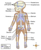
Divisions of the Skeleton
Axial and Appendicular Skeleton
Axial Skeleton (80 bones): Skull, vertebral column, and thoracic cage.
Appendicular Skeleton (126 bones): Limbs and girdles (pectoral and pelvic).
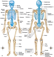
Spinal Curvatures
Normal Curves: Cervical, thoracic, lumbar, and sacral.
Abnormal Curves: Lordosis (swayback), kyphosis (hunchback), scoliosis (lateral curvature).
Thorax and Ribs
Ribs: 12 pairs (true, false, and floating ribs).
Sternum: Breastbone.
Thoracic Vertebrae: Form the posterior anchor for the ribs.
Upper and Lower Extremities
Upper Limb: Humerus, radius, ulna, carpals, metacarpals, phalanges.
Lower Limb: Femur, tibia, fibula, tarsals, metatarsals, phalanges, patella.
Pectoral Girdle: Scapula and clavicle.
Pelvic Girdle: Coxal bones, sacrum, coccyx.
Foot Structure
Arches: Medial and lateral longitudinal, and transverse (metatarsal) arches support body weight and provide leverage.
Sex and Age Differences in the Skeleton
Male vs. Female Skeleton
There are notable differences between male and female skeletons, especially in the pelvis, to accommodate childbirth in females.
Size: Male skeleton is generally larger.
Pelvis Shape: Male pelvis is deep and narrow; female pelvis is broad and shallow.
Pelvic Inlet: Wider in females.
Pubic Angle: Wider in females.
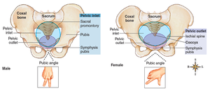
Age and Environmental Factors
Maturity: Skeleton matures around age 25.
Aging: Bone density decreases after age 50.
Nutrition and Stress: Affect bone health and structure.
Joints (Articulations)
Classification of Joints
Joints are classified by the degree of movement they allow and their structural characteristics.
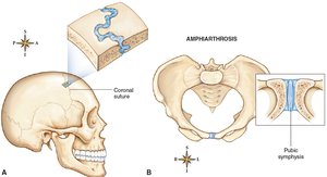
Synarthrosis: Immovable joints (e.g., skull sutures).
Amphiarthrosis: Slightly movable joints (e.g., pubic symphysis).
Diarthrosis (Synovial Joints): Freely movable joints (e.g., knee, shoulder).
Structure of a Diarthrotic (Synovial) Joint
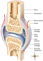
Joint Capsule and Ligaments: Hold bones together and permit movement.
Articular Cartilage: Covers bone ends, absorbs shock.
Synovial Membrane: Lines capsule, secretes lubricating fluid.
Joint Cavity: Space between articulating bones.
Types of Synovial Joints
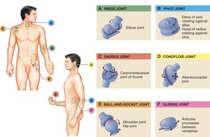
Ball-and-Socket: Shoulder, hip.
Hinge: Elbow, knee.
Pivot: Atlas and axis (neck).
Saddle: Thumb joint.
Gliding: Intercarpal joints.
Condyloid: Wrist joint.
Disorders of Bones and Joints
Tumors of Bone and Cartilage
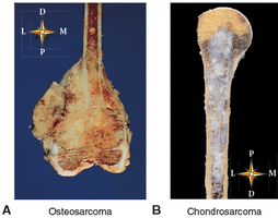
Osteosarcoma: Most common malignant bone tumor, often in distal femur, proximal tibia, or humerus.
Chondrosarcoma: Cancer of cartilage, second most common skeletal cancer.
Metabolic Bone Diseases
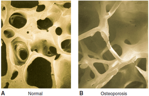
Osteoporosis: Loss of bone matrix and trabeculae, leading to fragile bones. Common in elderly, especially postmenopausal women.
Rickets: Vitamin D deficiency in children, causing soft bones and skeletal deformities.
Osteomalacia: Vitamin D deficiency in adults, leading to bone softening and increased fracture risk.
Paget Disease: Abnormal bone remodeling, resulting in deformed and fragile bones.
Osteogenesis Imperfecta: Genetic disorder causing brittle bones due to defective collagen.
Bone Infection
Osteomyelitis: Bacterial infection of bone, often requiring prolonged antibiotic therapy or surgery.
Bone Fractures
Open (Compound) Fracture: Bone pierces the skin.
Closed (Simple) Fracture: Bone does not pierce the skin.
Other Types: Complete, incomplete, linear, transverse, oblique.
Joint Disorders
Osteoarthritis (Degenerative Joint Disease): Most common noninflammatory joint disorder, characterized by joint pain and stiffness.
Dislocation/Subluxation: Bones in a joint are displaced from normal alignment.
Sprain: Injury to ligaments.
Strain: Injury to muscle or tendon.
Inflammatory Joint Disorders
Rheumatoid Arthritis: Systemic autoimmune disease causing chronic inflammation of joints and other tissues.
Gouty Arthritis: Caused by deposition of urate crystals in joints.
Infectious Arthritis: Joint inflammation due to infection (e.g., Lyme disease).
Additional info: This guide covers the essential structure, function, and pathology of the skeletal system, suitable for ANP college-level study and exam preparation.
