 Back
BackThe Skeletal System: Structure, Function, and Classification
Study Guide - Smart Notes
 Tailored notes based on your materials, expanded with key definitions, examples, and context.
The Skeletal System
Overview of the Skeletal System
The human skeletal system consists of approximately 206 bones and associated cartilages, forming the structural framework of the body. It is divided into two main parts: the axial skeleton and the appendicular skeleton. - Axial skeleton: Forms the body's longitudinal axis and encases body cavities, protecting vital organs. Includes the skull, vertebral column, and thoracic cage. - Appendicular skeleton: Comprises the bones of the pectoral and pelvic girdles, as well as the limbs, primarily suited for movement and muscle attachment.
Divisions of the Skeletal System
- Axial skeleton: Skull (22 bones: 8 cranial, 14 facial), vertebral column (33 bones), thoracic cage (12 pairs of ribs, sternum, part of vertebral column). - Appendicular skeleton: Pectoral girdle (clavicle, scapula), upper limb (humerus, radius, ulna, carpals, metacarpals, phalanges), pelvic girdle (pelvic bones, sacrum), lower limb (femur, tibia, fibula, tarsals, metatarsals, phalanges).
Bone Markings
Types and Functions of Bone Markings
Bone markings are surface features that reflect the structure-function relationship in bones. They are classified as depressions, openings, and projections. - Depressions: Pathways for blood vessels and nerves, or sites for articulations between bones. - Openings: Enclose delicate structures and allow passage through bones. - Projections: Sites for articulation or attachment of ligaments and tendons.
Table: Bone Markings
Bone Marking | Description | Example |
|---|---|---|
Facet | Shallow concave or convex surface where two bones articulate | Rib: Articular facet for articulation with transverse process |
Fossa (plural: fossae) | Indentation in a bone into which another structure fits | Humerus: Distal portion with olecranon fossa |
Fovea | Shallow pit | Femur: Fovea capitis |

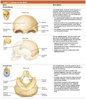
The Skull
Structure and Classification of Skull Bones
The skull consists of 22 bones organized into cranial and facial groups. Cranial bones protect the brain, while facial bones form the framework of the face. - Cranial bones: Four single bones (frontal, occipital, ethmoid, sphenoid) and two paired bones (temporal, parietal). - Facial bones: Six paired bones (maxillary, zygomatic, nasal, lacrimal, palatine, inferior nasal conchae) and two single bones (mandible, vomer). - Sutures: Immovable joints between skull bones, except for the mandible. - Sinuses: Air-filled, membrane-lined spaces in four bones surrounding the nasal cavity (paranasal sinuses).
Table: Bones of the Skull
Bone | Description |
|---|---|
Frontal Bone | Forms the forehead and superior part of the orbit. |
Parietal Bone | Forms the superior and lateral aspects of the skull. |
Occipital Bone | Forms the posterior and base of the skull. |
Temporal Bone | Forms the lateral and inferior aspects of the skull. |
Sphenoid Bone | Forms part of the base and sides of the skull. |
Ethmoid Bone | Forms part of the nasal cavity and the orbits. |
Maxilla | Forms the upper jaw and part of the orbit. |
Mandible | Forms the lower jaw. |
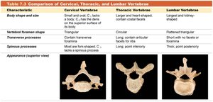
The Vertebral Column
Structure and Classification of Vertebrae
The vertebral column consists of 33 vertebrae, classified by region and function. It provides support, protects the spinal cord, and allows movement. - Cervical vertebrae (7): Located in the neck; characterized by transverse foramina. - Thoracic vertebrae (12): Articulate with ribs; heart-shaped bodies and long spinous processes. - Lumbar vertebrae (5): Largest and heaviest; kidney-shaped bodies and thick spinous processes. - Sacral vertebrae (5 fused): Form the sacrum; articulate with pelvic bones. - Coccygeal vertebrae (3–5 fused): Form the coccyx; most inferior part.
Table: Comparison of Cervical, Thoracic, and Lumbar Vertebrae
Characteristic | Cervical Vertebrae | Thoracic Vertebrae | Lumbar Vertebrae |
|---|---|---|---|
Body shape and size | Small and oval; C1 lacks a body, C2 has the dens | Larger; heart-shaped; contain costal facets | Largest and kidney-shaped |
Vertebral foramen shape | Triangular | Circular | Flattened triangular |
Transverse processes | Most are fork-shaped; C1 lacks a spinous process | Long; contain articular facets for ribs | Thick; point posteriorly |
Spinous processes | Short; fork-shaped | Long; point inferiorly | Thick; point posteriorly |
Appearance (superior view) |
|
|
|


Bone Markings and Skull Cavities
Bone Markings: Depressions, Facets, Fossae, and Foveae
Bone markings are essential for understanding how bones interact and provide sites for muscle attachment, articulation, and passage of nerves and blood vessels. - Depressions: Clefts of varying depth; found where bones meet other structures. - Facet: Shallow concave or convex surface for articulation. - Fossa: Indentation for another structure to fit. - Fovea: Shallow pit.
Skull Cavities
The skull contains several cavities that house sensory organs and provide passageways for nerves and blood vessels. - Orbits: Encases the eyeballs and associated structures. - Nasal cavity: First part of the respiratory tract; lined with mucous membranes. - Oral cavity: Surrounds teeth and tongue; first part of the digestive tract. - Other cavities: Contain organs for hearing and balance.
Study Boost: Mnemonics and Visual Analogies
Mnemonics for Remembering Bones
Mnemonics are useful for memorizing the names and locations of bones. - Pest Of 6: Parietal, Ethmoid, Sphenoid, Temporal, Occipital, Frontal (cranial bones) - Virgil Is Now Making My Pet Zebra Laugh: Vomer, Inferior nasal conchae, Nasal, Mandible, Maxillae, Palatine, Zygomatic, Lacrimal (facial bones) - Breakfast at 7, lunch at 12, dinner at 5: 7 cervical, 12 thoracic, 5 lumbar vertebrae
Visual Analogies
- Sphenoid bone: Looks like a bat with wings. - Ethmoid bone: Positioned like an iceberg, with only its tip visible. - Thoracic vertebra: Resembles the head of a giraffe. - Lumbar vertebra: Resembles the head of a moose.
Conclusion
The skeletal system is a complex structure that provides support, protection, and movement. Understanding the classification, structure, and function of bones, as well as their markings and relationships, is essential for students of anatomy and physiology. Additional info: Academic context and expanded explanations were added to ensure completeness and clarity for college-level study.
